Cell-secreted proteins, such as antibodies, hormones and neurotransmitters, play a crucial role in maintaining overall health and well-being. They are also key components in disease research and in developing diagnostic tools and personalized medicines. However, current methods for studying these proteins are limited to observing large groups of cells together, which makes it difficult to discern individual cell behaviors and differences.
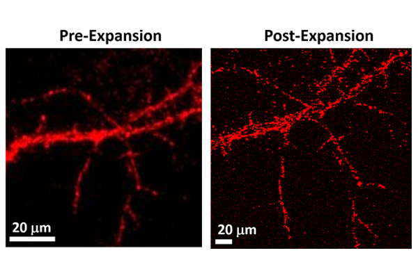
With a $450,000 grant from the National Science Foundation, Srikanth Singamaneni, the Lilyan & E. Lisle Hughes Professor in the McKelvey School of Engineering at Washington University in St. Louis, will develop a method called Plasmon-Enhanced Expansion FluoroSpot (PEEFS) to address these limitations. PEEFS combines a very bright fluorescent nanoparticle with expansion microscopy to image secreted proteins with high sensitivity and precision and accurately measure differences between cells.
The project represents a potentially transformative advance, particularly in immunology, oncology, stem cell biology and other life-science disciplines. With PEEFS, researchers will be able to image and quantify protein secretion at extremely high resolutions — down to the level of a single cell — revealing cell-to-cell variability and interactions and the spatial and temporal dynamics of cell-secreted proteins.
This story was originally published on the McKelvey School of Engineering website.