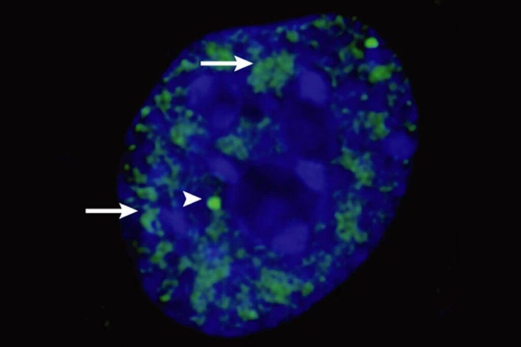
Genes are copied to make RNA transcripts, then the copies are translated into proteins. The transcripts, known as pre-messenger-RNA (pre-mRNA), are processed in the cellular nucleus to generate bona fide mRNAs that are then translated by ribosomes. This processing, known as splicing, happens in a cellular region known as nuclear speckles. Errors in splicing, which can happen as a function of aging, or cellular stress directly contribute to the onset and progress of various cancers and are associated with neurodegenerative disorders such as amyotrophic lateral sclerosis (ALS).
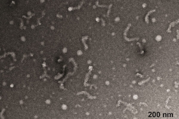
Speckles are thought to be biomolecular condensates, where DNA, RNA and proteins are organized into distinct molecular communities that form via phase separation. Rohit V. Pappu, the Gene K. Beare Distinguished Professor of Biomedical Engineering in the McKelvey School of Engineering at Washington University in St. Louis, and Min Kyung Shinn, a former postdoctoral researcher in his lab, investigated some of the physical features of nuclear speckles that were inconsistent with the type of uniform droplets one expected to see if they formed via liquid-liquid phase separation.
Shinn and Pappu set out to reconstitute the phase transitions of various RNA-binding proteins that belong to nuclear speckles. The results included finding unique structures of speckle-associated proteins, called microphases, as detailed in a recent publication in Cell. The team said the microphases seem to be the relevant targets for therapeutic intervention in specific cancers and in ALS because they have precise organizations of molecules.
Read more on the McKelvey Engineering website.