In vitro fertilization has been the primary method of addressing infertility for almost five decades, yet its live birth rate is under 40%. Selecting an embryo with the optimal potential could improve the success rate, but existing practices to take time-lapse images of developing embryos have been limited.
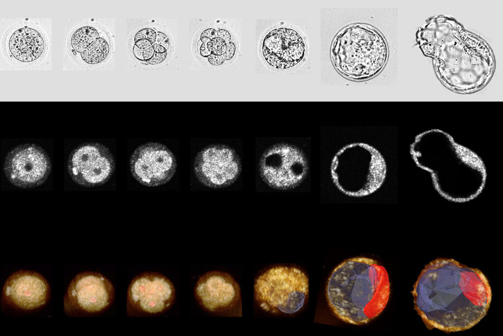
A team of researchers in the McKelvey School of Engineering at Washington University in St. Louis used dual-modality, 3D, time-lapse optical coherence microscopy (OCM) and brightfield (BF) imaging to monitor mouse embryo development and predict successful blastocyst formation. Results of this research, published in Communications Biology this spring, establish OCM as a viable way to choose high-quality embryos for transfer and improve the success rates of in vitro fertilization.
The innovative research was conducted in the lab of Chao Zhou, a professor of biomedical engineering and an internationally renowned expert in OCM and optical coherence tomography. Collaborators in the work include Fei Wang, a doctoral student in Zhou’s lab, and Ali Ahmady, an associate professor of obstetrics and gynecology at WashU Medicine.
Zhou will present this work at the American Society for Reproductive Medicine’s annual meeting in October. Read more on the McKelvey Engineering website.