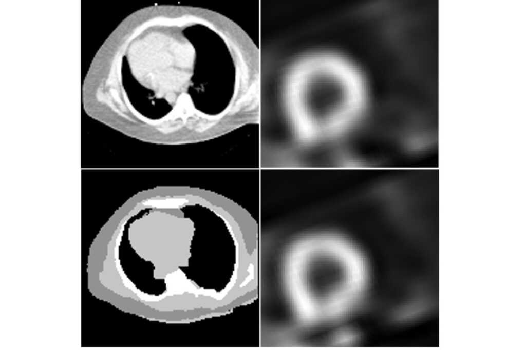
Coronary artery disease is a leading cause of death globally. A common tool to diagnose and monitor heart disease, myocardial perfusion imaging (MPI) by single photon emission computed tomography (SPECT), uses a radioactive tracer and special camera to provide detailed images of blood flow to the heart. It helps doctors detect coronary artery disease and other cardiovascular abnormalities. However, traditional SPECT imaging requires an additional CT scan to ensure accurate results, exposing patients to more radiation and increasing costs.
Now, a new deep-learning technique developed by researchers at Washington University in St. Louis — with collaborators from the Cleveland Clinic and the University of California, Santa Barbara — could transform the way heart health is monitored, making it safer and more accessible. The method, known as CTLESS, leverages deep learning to remove the CT requirement without compromising diagnostic accuracy. The project, led by Abhinav Jha, an associate professor of biomedical engineering at the McKelvey School of Engineering and of radiology at WashU Medicine Mallinckrodt institute of Radiology, was published online Nov. 25 in IEEE Transactions on Medical Imaging.
The researchers’ next steps are to validate this method while also working to make the technology more available to rural community hospitals. Their cost-saving technique is particularly significant for cases where access to such scans may be limited, such as in rural or otherwise resource-limited communities, Jha said.
Read more on the McKelvey Engineering website.