Biomedical engineer Abhinav Jha, an assistant professor at the McKelvey School of Engineering and of radiology at the School of Medicine, both at Washington University in St. Louis, has published two papers recently related to improving imaging methods for medical applications.
Jha has long advocated that artificial intelligence (AI) tools used in medical applications for image processing need to be evaluated based on clinical tasks, not visual appeal.
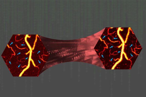
In a study published in IEEE Transactions on Radiation and Plasma Medical Sciences, Jha and his collaborators developed a tool that demonstrates the potential to improve performance on clinical tasks. The tool has been developed in the context of denoising myocardial perfusion imaging (MPI) single-photon emission computed tomography (SPECT) images, which help evaluate blood flow to the heart muscle.
Read more on the McKelvey School of Engineering website.
Separately, Jha and his collaborators developed a new imaging method for personalized cancer treatment. Recently published results from a computer-based clinical trial indicate that the method may be reproducible in humans and merits additional clinical testing.
In a study published in the Journal of Nuclear Medicine, Jha and collaborators evaluated the effectiveness of a specialized single-photon emission computed tomography (LC-QSPECT) imaging method to estimate doses of alpha-particle-emitting radiopharmaceutical therapy (a-RPT).
Read more about that work on the McKelvey Engineering website.