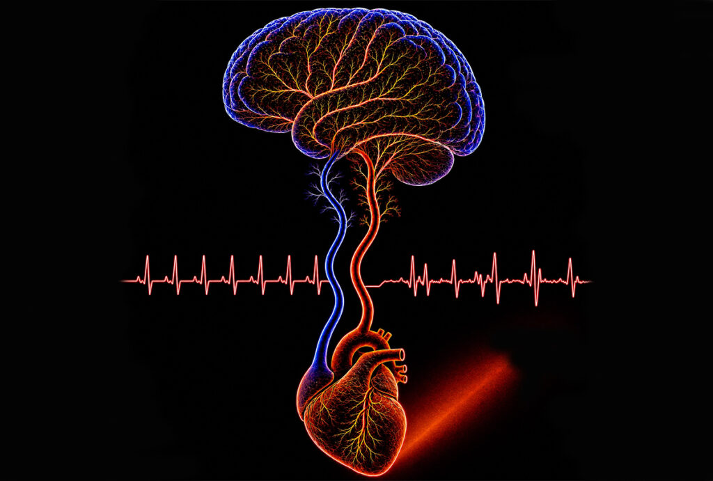
An irregular heartbeat, or arrhythmia, leads to inefficient pumping of blood by the heart, which then prevents blood and oxygen from getting to the body’s other organs. When blood and oxygen flow poorly to the brain, the risk of stroke and cognitive decline increases.
A team of researchers based at Washington University in St. Louis used cardiac optogenetics to noninvasively study arrhythmia and its impact on the brain. Using highly sensitive imaging in a mouse model, they found that arrhythmia in a mouse heart alters oxygen concentration in the brain during and after arrhythmia.
Results of the research are published in Science Advances June 3.
Chao Zhou, a professor of biomedical engineering at WashU McKelvey Engineering; Abby Matt, a graduate student in Zhou’s lab; and Marcello Magri Amaral, a former visiting researcher from Universidade Brasil, led a team using optogenetics — the combination of genetic engineering and light to control neurons or cardiac cells. They produced a stimulation in genetically modified mice. By shining a red light on the skin over the mouse heart, they could safely change the pacing of the heartbeat up to 175% of resting heart rate to create arrhythmia on demand. Afterward, the heart rate returned to its initial resting rate.
In their experiments, they used several frequencies, ranging from a below-resting heart rate of 6 Hz to 14 Hz, which is well above resting rate, while recording the electrocardiogram signal during rest, light pacing and recovery periods. The largest changes in the brain took place at 6 Hz and 14 Hz. Across the range of frequencies, changes in blood and oxygen concentration were in proportion with the discrepancy between the resting heart rate and the pacing frequency.
“With this model, we showed that we reduced the cardiac output, which is the volume of blood per second coming out from the heart, and then from this, we extended it to study a very highly perfused organ, the brain,” Matt said.
The team worked with the group of Adam Bauer, an associate professor of radiology at WashU Medicine Mallinckrodt Institute of Radiology, and of biomedical engineering at McKelvey Engineering.
“We can decrease the concentration of oxygenated hemoglobin and increase the amount of deoxygenated hemoglobin in the brain,” Matt said. “So, starting from a heart scale, we’re able to override rhythm. And we can use that to study highly perfused organs and how these are disrupted on a very short timescale.”
Optogenetics began at Stanford University in 2005 focusing primarily on neuroscience. Zhou first published his work in cardiac optogenetics in Science Advances in 2015 while at Lehigh University, where he used the method to change the heartbeat in the fruit fly. His team later made additional fruit fly models that are responsive to different colors of light.
Traditional methods of heart pacing are invasive and technically complex, requiring direct implantation of LED sources on the heart surface or high power, which risks burning the skin.
“Our advantage with using light is that it’s noninvasive and doesn’t require a wire or an electrical lead to do pacing,” Matt said. “Instead, the effect is only confined to where our light-sensitive proteins are expressed, so cardiac optogenetics is more targeted and gives us more control.”
Future work may combine optical intrinsic signal imaging with other methods that measure blood flow and oxygen saturation to give a wider perspective of how arrhythmia-induced blood flow changes affect the brain and other organs. While the team showed the method is effective in mice, they say it could be extended to other cardiac optogenetic models that may have greater potential for translational medicine.
Amaral MM, Matt A, Schloss KH, Wang F, Gracheva E, Wang Y, Liang H, Bice A, Ding J, Kovacs A, Weinheimer C, Diwan A, Cui J, Rentschler S, Nerbonne J, Zemlin C, Bauer AQ, Zhou C. Non-invasive optogenetic induction of cardiac arrhythmias alters systemic hemodynamics in mice. Science Advances, June 3, 2026. DOI: 10.1126/sciadv.aeb1092
Support for this research is provided by Washington University in St. Louis; the National Institutes of Health (R01-EB025209, R21-EB032684, R01-HL156265, R01NS10287005, R01NS12632601, and RF1AG07950301); the National Science Foundation Graduate Research Fellowship Program (GRFP); and American Heart Association (25PRE1365124).