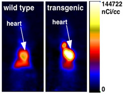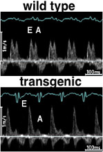
In mice whose heart muscles take up high amounts of fat, the heart fills abnormally after each contraction, researchers at Washington University School of Medicine in St. Louis have found. This condition is consistent with the first stage of heart dysfunction in human diabetics.
Heart disease is the leading cause of death among the more than 13 million diabetics in the United States. Clinical studies suggest impairment of the diastolic or filling phase of the cardiac cycle is the first stage of a progression that leads to more widespread heart malfunction in patients with diabetes.
“For the first time we’ve been able to reproduce many aspects of what you see very early on in the hearts of individuals with diabetes by altering fat metabolism in the heart,” says Jean Schaffer, M.D., associate professor of medicine and of molecular biology and pharmacology. “If these mice can lead us to ways to intervene at this stage, we might be able to help prevent diabetic cardiomyopathy.”
The study was reported in the February issue of Circulation Research.
The mice used in the study were engineered so that their cardiac muscles produce eight times the normal amount of a protein that pulls fat into cells. The heart muscle responds by increasing its rate of fat burning. But the uptake of fat exceeds the ability of the cells to adjust their metabolism, and the cells accumulate two times more fat than normal.

Many diabetics have high levels of fat in their blood, which can contribute to abnormal fat metabolism and the accumulation of fat in heart tissue. Precisely how this leads to the symptoms of heart disease seen among diabetics has not yet been clarified.
“These mice allow us to isolate and study in detail the effect of fat on heart muscle cells, independent of any types of global metabolic effects caused by abnormal blood levels of fats,” says Schaffer, a cardiologist at Barnes-Jewish Hospital.
The research team performed echocardiograms or sound wave imaging of the heart and performed cardiac catheterizations to measure the pressures within the mouse hearts during the cardiac cycle. The results of these studies indicated that the ability of the heart to relax after contracting was impaired. In other words, the hearts were stiff or noncompliant.
When the mice’s heart tissue was examined microscopically, there was evidence of fibrosis, an increased deposition of fibers surrounding the heart cells.
“Additional fibrous material such as this can change the elasticity of cardiac tissue,” Schaffer says. “So that may contribute to impaired relaxation.”
The hearts also showed significant prolongation of the time between electrical activation and inactivation of the ventricles. This particular change in the electrical properties of the heart appears frequently among diabetics and is a predictor of cardiac mortality.

Previously, the research team had developed other transgenic mice with increased amounts of other proteins that bring fats into cells. These earlier mouse models demonstrated different heart problems, including defects in pumping action, rather than in the filling phase. In addition, those mice often died young, while the mice used in the present studies had a normal lifespan.
“The difference between the mouse models we’ve developed tells us that how fats enter heart muscle cells may determine how the cells cope with it,” Schaffer says. “We will continue investigating the mice to clarify the link between excess fat, fat metabolism, and filling problems in the heart. We also will begin to test what kinds of therapeutic interventions decrease the apparent toxicity of fat on the heart and help remedy the heart defects shown in the mice.”
Chiu H-C, Kovacs A, Blanton RM, Han X, Courtious M, Weinheimer CJ, Yamada KA, Brunet S, Xu H, Nerbonne JM, Welch MJ, Fettig NM, Sharp TL, Sambandam N, Olson KM, Ory DS, Schaffer JE. Transgenic expression of fatty acid transport protein 1 in the heart causes lipotoxic cardiomyopathy. Circulation Research Feb. 2005;96:225-233.
Funding from the National Institutes of Health and the American Heart Association supported this research.
Washington University School of Medicine’s full-time and volunteer faculty physicians also are the medical staff of Barnes-Jewish and St. Louis Children’s hospitals. The School of Medicine is one of the leading medical research, teaching and patient care institutions in the nation, currently ranked third in the nation by U.S. News & World Report. Through its affiliations with Barnes-Jewish and St. Louis Children’s hospitals, the School of Medicine is linked to BJC HealthCare.