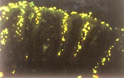
The bacterium that causes ulcers and contributes to stomach cancers uses a clever interaction between two genes to randomly tighten and loosen its grip on the stomach, according to a study by researchers at Washington University School of Medicine in St. Louis and Umeå University in Sweden.
Helicobacter pylori often binds tightly to cells of the stomach lining to feed, but the newly identified interaction ensures that a small reservoir of bacteria are always more loosely connected. This reservoir is much more likely to survive if the host mounts a strong immune response.
“Basically, if you’re holding onto someone’s T-shirt and they start punching you hard, you’d like to be able to let go,” jokes Douglas Berg, Ph.D., Alumni Professor of Molecular Microbiology and an author of the study. “Any savvy bacteria are going to want to be able to do the same.”
New insights into how H. pylori sticks to and then releases from the stomach wall will advance efforts to design better drugs and vaccines against the bacterium, which is estimated to be present in more than half of the world’s population.
Most H. pylori infections in the U.S. and other industrialized nations can be treated with antibiotics, but treatments are too costly for many sufferers in underdeveloped nations, where the bacteria’s pervasiveness and poor sanitation significantly increase the risk of repeat infections. In addition, resistance to standard drug therapies is a major problem in these countries.
The study appears in the online edition of Proceedings of the National Academy of Sciences. It will appear in print in the journal on November 30.

Researchers at Umeå University led by Anna Arnqvist, Ph.D., associate professor of molecular biology and medical biochemistry and a former Washington University predoctoral student, studied a Swedish strain of H. pylori. They focused on BabA, a protein that binds to Lewis B antigen receptor, a carbohydrate structure on the surface of stomach cells. Because they help organisms stick to particular targets in a glue-like fashion, BabA and proteins like it are collectively known as adhesins.
One of the Swedish strain’s two copies of the gene for BabA is “silent,” or blocked from use by damage in a region of DNA normally involved in the gene’s activation. The second copy is missing an essential portion of DNA, making it completely nonfunctional.
“This suggested that the strain doesn’t make BabA protein at all, making it equivalent in that regard to about one-third of all the other clinical isolates of H. pylori scientists have studied,” Berg says.
Given that the strain didn’t appear to produce BabA protein, the bacteria should have been unable to get a grip on the Lewis B receptor. However, scientists found that a small minority of the bacteria still stuck very tightly to Lewis B.
Researchers then determined that this resulted from the bacteria recombining DNA from the silent BabA gene and DNA from the gene for a similar protein, BabB. Scientists aren’t sure what, if anything, BabB sticks to, but they do know that its similarities to BabA include biochemical “hooks” at the beginning and end of the protein. These hooks anchor the proteins in the bacteria’s cell wall.
The BabA gene’s middle section encodes the glue that makes the protein stick. The rare bacteria that could grip Lewis B had spliced that middle section from the silent BabA gene into BabB, providing themselves with the equivalent of a working BabA gene and its protein product.
BabA-BabB gene recombination is relatively rare because characteristics of the segments of DNA being combined make them technically difficult for the bacteria to splice together. In addition, the BabB gene has a built-in genetic feature that allows the gene to turn on and off irregularly as the bacteria reproduce.
“The BabB gene has a highly repetitive section that has a tendency to slip when the bacteria copies its DNA prior to cell division,” Berg explains. “These slips can introduce extra repeats or delete them, shifting how the gene is translated from DNA to protein in a way that’s likely to halt protein synthesis by introducing a premature stop signal.”
The net result, according to Berg, is that the bacteria’s ability to stick to the Lewis B receptor is metastable—in every generation, a small number of the new bacteria will switch from a tight grip to no grip, or vice-versa.
“This metastability is likely an important component of the bacteria’s ability to adapt to host immune system responses,” Berg says.
Berg and his Swedish colleagues are currently working to better understand BabB, investigating, among other things, whether the gene has a role to play on its own as a producer of a bacterial adhesin or only acts as a random enabler of BabA.
Among the approximately 30 H. pylori surface proteins so far known to scientists, researchers have found other pairs of closely related genes. Included in these pairs are other genes that code for surface adhesins.
“We also will take a close look at some of these pairs,” Berg says. “We’re eager to find out whether they contain variations of this special regulatory system in BabA and BabB, or whether they control the strength and specificity of H. pylori adherence in other ways.”
Bäckström A, Lundberg C, Kersulyte D, Berg DE, Borén T, and Arnqvist A. Metastability of Helicobacter pylori genes and dynamics in Lewis B antigen binding. Proceedings of the National Academy of Sciences, November 30, 2004, 16923-16928.
Funding from the J.C. Kempe Memorial Foundation, the B von Kantzows Foundation, the Tore Nilsson Medical Research Foundation, the Magnus Bergwall Research Foundation, the Medical Faculty of Umeå University, the Kempestiftelserna, the Swedish Medical Research Council, the Swedish Cancer Society, the Umeå University-Washington University exchange program, and the National Institutes of Health.
Washington University School of Medicine’s full-time and volunteer faculty physicians also are the medical staff of Barnes-Jewish and St. Louis Children’s hospitals. The School of Medicine is one of the leading medical research, teaching and patient care institutions in the nation, currently ranked second in the nation by U.S. News & World Report. Through its affiliations with Barnes-Jewish and St. Louis Children’s hospitals, the School of Medicine is linked to BJC HealthCare.