School of Medicine pediatric plastic and reconstructive surgeons and students have launched a unique database of 3-D craniofacial images of more than 1,200 children that will help researchers study the form and growth of the head and face.
The database, called Craniobank (craniobank.wustl.edu), is the first free and searchable online database of images of children from infants to age 18 of various ethnic backgrounds without craniofacial disorders. The images will be available to researchers in a variety of fields, including anthropology, psychiatry, plastic and reconstructive surgery, neurology and dentistry, to lead to earlier diagnoses and a better understanding of craniofacial conditions.

Lipira
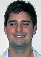
Spearheading the endeavor was Angelo Lipira, now a fourth-year medical student. Lipira developed the database during a yearlong Doris Duke Clinical Research Fellowship with Alex A. Kane, M.D., the Dr. Joseph B. Kimbrough Chair for Pediatric Dentistry and a craniofacial surgeon at St. Louis Children’s Hospital, and an information technology team from the Department of Surgery.
Craniobank allows researchers to perform searches by age, gender, ethnicity, facial expression and hand preference to assemble a group of healthy subjects for studies.
“Our goal is for other investigators to be able to download large sets of data to be used in studies of craniofacial form and structure,” Lipira said. “This would include studies of pathological conditions where our normative data could serve as controls or studies of normal craniofacial growth and development.”
Although Kane and his colleagues have built an internal archive of patients they treat for a variety of disorders of the head and face, such as birth defects, injuries and burns, there was no existing online database of so-called normal head and face shapes for comparison.
Before Craniobank, the “bible” of normative data was developed by Leslie Farkas, M.D., a Canadian physician who painstakingly measured angles and distances between points on the face and head and listed measurements by gender and age in a book titled “Anthropometry of the Head and Face.”
“That data is great, but it’s not really reproducible,” said Kane, associate professor of plastic and reconstructive surgery and director of the Cleft Palate and Craniofacial Institute at St. Louis Children’s Hospital. “That’s the beauty of Craniobank — it’s there digitally. If someone wants to use this data for a purpose that we can’t anticipate, they can download it and use it for any mathematical or analytical technique they desire and apply it to that data.”
Lipira visited several area pediatricians’ offices to acquire images of children, with parental permission, using a sophisticated 3-D camera system called digital stereophotogrammetry. The system uses 12 cameras to take simple photographs of the head from four different angles and takes less than one second. The photos are then calibrated using software to reconstruct a high-resolution image.
The stereophotogrammetry equipment is used daily in the Cleft Palate and Craniofacial Institute at St. Louis Children’s Hospital to acquire images of all patients before and after surgery and to evaluate their growth over time.
Researchers must register to access the data and then provide approval from their institution’s Institutional Review Board or equivalent to use it. Nonregistered users can access the database but will not see images of the children.
Kane credits Lipira for his determination in getting the project done.
“He was incredibly dynamic in trying to get what he needed to get in a small window of time given the scope of the project,” Kane said. “I have to give all the credit to Angelo, who really took it and ran in a way that I couldn’t have anticipated.”
The researchers also plan to acquire more images of children from underrepresented ethnic groups. In the future, Kane would like to add DNA information with each image to allow for genetic studies.