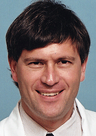
Russell N. Van Gelder, M.D., Ph.D., is the new Bernard Becker Professor of Ophthalmology and Visual Sciences. Larry J. Shapiro, M.D., executive vice chancellor for medical affairs and dean of the School of Medicine, announced the appointment.

“As an outstanding physician-scientist making important contributions to our understanding of the visual system, Russ Van Gelder is a superb candidate for this recognition,” Shapiro said.
“This professorship also reminds us of the legacy and generosity of Dr. Becker. His research and record as a teacher and administrator are unsurpassed. Bernie is unequalled as a supporter of research and education in general, and of Washington University in particular.”
Becker, M.D., has more than 50 years of involvement with education, the arts and social causes in St. Louis. This professorship is one of two endowed chairs in the Department of Ophthalmology and Visual Sciences that were originally established in 1983 in recognition of the service and leadership of Becker, professor emeritus and former head of ophthalmology.
“Dr. Becker is a principal figure in the history of our department, and the professorships endowed in his name continue to advance vision research,” said Michael A. Kass, M.D., head of the department and a former resident under Becker. “Russ Van Gelder’s important work is providing both insight into visual disorders and non-visual systems regulated by cells in the eye.”
Van Gelder came to the School of Medicine in 1995 as a resident in the ophthalmology and visual sciences department. He later became an instructor and postdoctoral fellow, studying eye inflammation. He joined the faculty full time as assistant professor in 1999, and a year later became assistant professor of molecular biology and pharmacology.
A specialist with the Barnes Retina Institute, Van Gelder also directs the Uveitis Service for the ophthalmology and visual sciences department. In the laboratory, he concentrates most of his attention on non-visual pathways in the eye and their relationship to circadian rhythms. He has characterized pathways that let the eye sense light without seeing it.
Even in some completely blind eyes, these non-visual pathways still function to control the body’s circadian clock, using a retinal protein called melanopsin to operate an internal “light meter” that helps distinguish day from night even if visual processes don’t operate normally.
“Cells called intrinsically photosensitive retinal ganglion cells, or ipRGCs, can detect brightness, and they’re extremely good at it,” Van Gelder said. “You could make a good light meter for a camera out of these cells because they are consistent in their response to brightness, and that’s completely different from the way rods and cones behave in the retina. Those visual cells can’t detect brightness very well. They detect contrast, sensitivity and motion.”
Studying these populations of ganglion cells, Van Gelder also has found they require melanopsin to sense and react to pulses of light and that there are three distinct populations of ipRGCs in the retina, with each cell type reacting to light differently. He’s also learned that both animals and people with damaged ipRGCs tend to have problems with their circadian clocks and their sleep/wake cycles.
“Taken together, our results have led us to the unexpected conclusion that eye disease can be a risk factor for sleep disorders, and because these retinal ganglion cells are part of the optic nerve, optic nerve health strongly influences risk of sleep disorders,” Van Gelder said.