Scientists at Washington University School of Medicine in St. Louis have developed a method to predict whether melanoma of the eye will spread to the liver, where it quickly turns deadly. They also believe the molecular screening test may one day help determine the prognosis of patients with some types of skin melanoma.

J. William Harbour, M.D., the Paul A. Cibis Distinguished Professor of Ophthalmology and Visual Sciences and associate professor of cell biology and molecular oncology, reported on the screening test today at the American Academy of Cancer Research meeting in Chicago.
“About half of patients with ocular melanoma develop metastasis in the liver,” says Harbour, who directs the ocular oncology service at the School of Medicine. “Ocular melanoma has a strong propensity to spread to the liver, and when it does, it usually leads to death within a very short time.”
Doctors have known for many years that patient age, tumor size and location and shape of tumor cells all could help predict whether ocular melanoma was likely to spread. But none of those factors were accurate enough to influence treatment decisions in individual patients.
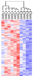
Now Harbour and colleagues have found that a particular molecular signature — that is, the pattern of activation of a group of genes in the tumor cells — accurately predicts risk for metastasis. Rather than analyzing a single protein or molecular factor, the test looks at how several factors work together.
“We were attempting to analyze these patterns the same way that our brain’s work to recognize a face to tell whether a person is ‘John’ or ‘Jane,'” Harbour explains. “We don’t just look at a nose or an eye. We look at the whole face. And in this project we used computer software to look at many, many features of the tumor simultaneously.”
Harbour’s efforts identified two classes of tumors with distinct molecular signatures. One signature, called class 1, carries a low risk of metastasis – less than 90%. Tumors with a class 2 signature have a greater than 90 percent chance of spreading to the liver.
When he first identified the molecular signatures, Harbour was testing tumor tissue taken from cancerous eyes that had been surgically removed. But only about 10 percent of ocular melanoma patients have such drastic surgery. Most have tumors that are small enough to be treated with radiation therapy.
Because the eye remains intact in the vast majority of patients, Harbour’s team needed to learn whether it is possible to run the molecular test on tumor samples gathered with a fine needle biopsy.
And the answer was ‘yes.’ “Even with the small amount of tumor tissue you get from a needle biopsy, the accuracy of the test is comparable to what we found when we had the entire tumor to work with,” he says.
Harbour’s molecular test can detect both whether a tumor is likely to spread to the liver and how fast. Some tumors tend to spread quickly while others take several years.
The researchers found two sub-groups of class 2 tumors, which differ mainly in a particular region of chromosome 8. One of these subgroups has lost a section of DNA called the short arm of chromosome 8, what’s known as chromosome 8p.
“If a patient has a class 2 tumor, and they have lost chromosome 8p, then that person is at high risk for spread of the cancer into the liver and at high risk that it will occur rapidly,” he says.

Knowing that the cancer is likely to spread quickly from the eye to the liver may allow for earlier, preventive treatments in high-risk patients. Harbour says at the very least, a person with a class 2 molecular signature should receive more frequent and more intensive surveillance to monitor the spread of the cancer. Many also may be candidates for pre-emptive therapy of some kind.
Melanoma of the eye is relatively rare, diagnosed in only about seven people per million each year in the United States or about 2,000 cases annually. But Harbour says that over a million additional individuals in the United States have one or more pigmented tumors in the eye that are too small to be called melanomas. Known as a nevus, or mole, the tiny, pigmented tumors can eventually develop into a melanoma. Harbour predicts the information gained from the genetic signatures in ocular melanomas may one day help to predict which of the small moles will turn into melanomas, thus allowing them to be treated earlier to reduce the chance of metastasis.
About half of all patients with eye melanoma have their tumors detected during routine eye exams, before they begin to affect vision. The other half experience blurred vision, see flashing lights and distortions or have defects in the visual field that usually involve blank spots in their peripheral vision. Most of the time, those symptoms won’t mean there’s an ocular melanoma, Harbour says, but they should be examined by an eye doctor.
Unlike skin melanomas, these eye tumors don’t seem to be related to ultraviolet light exposure, but Harbour says he is learning that the molecular signatures of eye melanomas can be remarkably similar to those seen in some forms of more common skin melanoma.
“When we look at skin melanomas, we see similar molecular signatures that distinguish the lower-grade, horizontal growth pattern from the higher-grade, vertical growth pattern,” he says. “So we are working very closely with other researchers at Washington University who study skin melanoma to see whether this test might be useful in predicting whether some patients with skin cancer might be at increased risk for metastasis.”
Habour JW, Onken MD, Worley LA. Molecular prediction of time to metastasis from ocular melanoma fine needle aspirates. Presented to the American Academy of Cancer Research, Sept. 13, 2006.
This research was supported by grants and awards from the National Eye Institute and the National Cancer Institute of the National Institutes of Health, the Tumori Foundation, the Horncrest Foundation, the Barnes-Jewish Hospital Foundation, Research to Prevent Blindness, Inc. and the Macula Society Retina Research Foundation/Mills and Margaret Cox Endowment Fund.
Washington University School of Medicine’s full-time and volunteer faculty physicians also are the medical staff of Barnes-Jewish and St. Louis Children’s hospitals. The School of Medicine is one of the leading medical research, teaching and patient care institutions in the nation, currently ranked fourth in the nation by U.S. News & World Report. Through its affiliations with Barnes-Jewish and St. Louis Children’s hospitals, the School of Medicine is linked to BJC HealthCare.