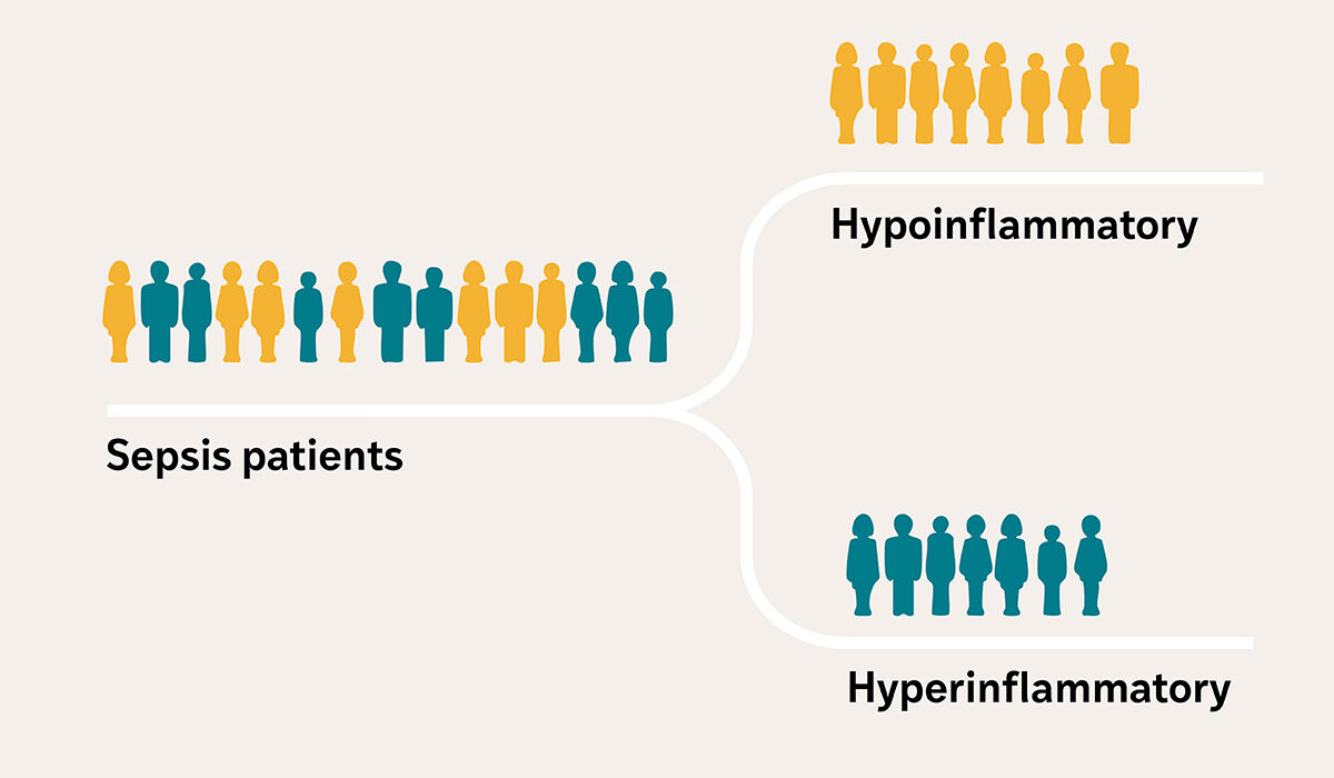
Critically ill patients with sepsis — a condition in which the body’s response to an infection spirals out of control and can lead to organ damage — often arrive at the intensive care unit (ICU) with similar symptoms, such as fever, low blood pressure and kidney failure, among others. But researchers at Washington University School of Medicine in St. Louis have revealed that sepsis patients actually fall into two distinct profiles, based on biological clues in their blood: one with severe inflammation and organ damage, and the other with a less severe response.
Targeting treatments to these biological profiles could help doctors treat people with sepsis more effectively, according to Pratik Sinha, MBChB, PhD, an assistant professor in the Department of Anesthesiology at WashU Medicine. In particular, therapies that were previously deemed unsuccessful may prove to be effective for patients with the severe inflammation profile. But there aren’t clinical tools to accurately and quickly identify these subgroups, which have also been found among patients with other critical illnesses, such as acute respiratory distress syndrome, a condition often caused by sepsis that results in severe lung failure.
A research team led by Sinha has received a $4.87 million grant from the U.S. Department of Defense to develop a clinical test and handheld device to measure the amount of two biological markers in the blood that can help physicians quickly group patients with sepsis into either a high-risk hyperinflammatory profile or a less severe hypoinflammatory profile.
“Developing a precise test and an affordable device to quickly identify patients with the high-risk hyperinflammatory profile is crucial for delivering targeted treatments and saving lives in critical situations when time matters,” said Sinha. “A highly portable device will enable deployment in active military and combat zones, where trauma increases the risk of infection and sepsis.”
Patient subgroups may advance sepsis care
Each year in the U.S. alone, at least 1.7 million people develop sepsis, and roughly 21% of them die. Included in that statistic are military personnel with serious infections due to trauma inflicted in combat. The mortality rate increases to around 50% in patients with the hyperinflammatory profile of sepsis. Treatment includes antibiotics to fight the infection and supportive therapy for organs that have endured damage, but this approach misses an important piece, according to Sinha.
“Current treatments don’t address the body’s dysregulated response to the infection,” he said. “The challenge in delivering effective treatments is the variability in the severity of the biological response among patients with similar symptoms.”
An earlier study of 3,000 people with critical illness by Sinha and his colleagues, published in The Lancet Respiratory Medicine, found that the blood levels of two inflammatory cytokines — interleukin-8 (IL-8) and soluble tumor necrosis factor receptor 1 (sTNFR-1) — were reliable indicators of whether the person has a high or low inflammatory response. Inflammatory cytokines are small molecules that cells use to communicate and that can either promote or reduce inflammation.
Many treatments tested in clinical trials on a collective sepsis patient population have been deemed ineffective, Sinha explained. But when the researchers performed a secondary analysis using data from two previously conducted clinical trials and categorizing patients into the two inflammatory profiles, they found that treatment efficacy differed between the two groups. Separating patients into subgroups to enrich clinical trials with patients with similar biological responses could potentially unlock new treatments and repurpose previously failed treatments to improve outcomes in sepsis and other critical illnesses.
Precise, portable, affordable
Supported by this new funding, Sinha is collaborating with Srikanth Singamaneni, PhD, the Lilyan & E. Lisle Hughes Professor at the McKelvey School of Engineering at WashU, to develop a rapid clinical test to capture IL-8 and sTNFR-1 from a few drops of plasma — the liquid component of blood that remains after removing blood cells — and a portable, handheld device to measure the amount of the captured proteins.
This so-called lateral flow test — similar to a COVID-19 rapid antigen test — would be the first to detect IL-8 and sTNFR-1 on a single test strip. Unlike an at-home COVID-19 test, which only indicates the presence of the SARS-CoV-2 virus, the researchers’ test incorporates small particles that glow, also referred to as plasmonic fluors, to measure the amount of each inflammatory protein captured on the strip. The researchers will build a device with a camera to detect the glowing particles, and a computer model will quantify the levels to categorize patients into the two subgroups.
“The biomarkers we are trying to detect are found in very small concentrations in the blood,” said Singamaneni, who developed the lateral flow system that incorporates the plasmonic fluors. The plasmonic fluor technology has been patented and licensed to Brightest Bio, a WashU startup formerly called Auragent BioSciences, by WashU’s Office of Technology Management. “Incorporating the fluorescent nanoparticles into the test renders it extremely sensitive, allowing us to not only detect but also accurately measure these biomarkers,” Singamaneni added. “Its sensitivity sets our test apart from other lateral flow assays and is crucial for classifying patients into groups.”
The researchers are developing the test and device to be affordable, accurate and fast. Its compact and portable design will allow it to be deployed directly in a variety of critical care settings, including military bases, combat zones and rural hospitals.
“This tool has the potential to reach patients at the bedside, offering personalized care based on each individual’s specific biological responses,” said Sinha. “We hope to fill a need in critical care that will help bring more effective treatments to more patients.”
The U.S. Army Medical Research Acquisition Activity, 808 Schreider Street, Fort Detrick MD 21702-5014 is the awarding and administering acquisition office. This work was supported by The Assistant Secretary of Defense for Health Affairs endorsed by the Department of Defense, in the amount of $4,876,076.00 through the Peer Reviewed Medical Research Program under Award Number HT9425-25-1-0825. Opinions, interpretations, conclusions, and recommendations are those of the author(s) and are not necessarily endorsed by the Department of Defense.
About Washington University School of Medicine
WashU Medicine is a global leader in academic medicine, including biomedical research, patient care and educational programs with 2,900 faculty. Its National Institutes of Health (NIH) research funding portfolio is the second largest among U.S. medical schools and has grown 83% since 2016. Together with institutional investment, WashU Medicine commits well over $1 billion annually to basic and clinical research innovation and training. Its faculty practice is consistently within the top five in the country, with more than 1,900 faculty physicians practicing at 130 locations. WashU Medicine physicians exclusively staff Barnes-Jewish and St. Louis Children’s hospitals — the academic hospitals of BJC HealthCare — and treat patients at BJC’s community hospitals in our region. WashU Medicine has a storied history in MD/PhD training, recently dedicated $100 million to scholarships and curriculum renewal for its medical students, and is home to top-notch training programs in every medical subspecialty as well as physical therapy, occupational therapy, and audiology and communications sciences.
Originally published on the WashU Medicine website