While early detection of breast cancer is critical, early prediction of how well the neoadjuvant chemotherapy treatment before surgery is working also may provide a window of opportunity when treatment could be altered and have a big impact on the patient’s quality of life.

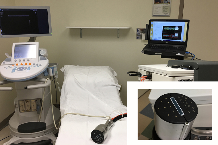
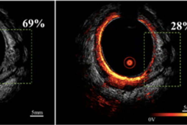
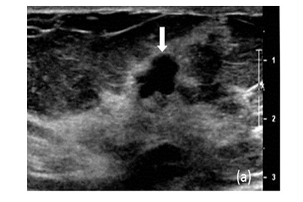
An interdisciplinary team of researchers at Washington University in St. Louis has found that combining data from tumor biomarkers, ultrasound and ultrasound-guided diffuse optical tomography (DOT) after a patient’s first cycle of pre-surgical neoadjuvant chemotherapy provided a highly accurate prediction of how the tumor was responding to the treatment. The results from a clinical trial at Washington University School of Medicine and Barnes-Jewish Hospital were published online in Breast Cancer Research and Treatment May 10.
Quing Zhu, professor of biomedical engineering at the McKelvey School of Engineering and of radiology at the School of Medicine, led a team of engineers and radiologists in the three-year clinical trial, which involved patients with various types of breast cancer, including triple-negative breast cancer, human epidermal growth factor receptor 2 (HER2+), and estrogen receptor-positive/human epidermal growth factor 2 negative (ER+/HER2-).
By conducting imaging after the first cycle of neoadjuvant chemotherapy, which ranges from two to three weeks depending on the treatment regimen used, the prediction model based on tumor biomarkers and imaging parameters can predict how well the cancer is responding, Zhu said.
Read more on the engineering website.


