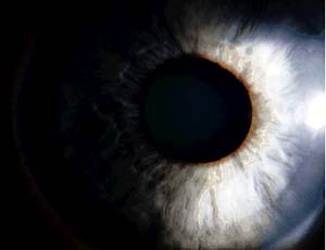
For years, science teachers have explained to their students that the eye is like a camera: The lens allows light to enter the eye, and the retina — like film — processes images and allows us to see.
In spite of how well that metaphor works, it’s probably time for an update. For one thing, as we plunge deeper into the digital age, fewer children will know what film is (c.f. record albums, eight-track tapes and black rotary phones). There’s also a key piece missing in the comparison. Most cameras also have light meters, and recent research suggests that the eye’s “light meter” is involved in more than just vision.

A light meter cannot form images, but it can determine how bright the environment is. In a camera, that information helps the photographer determine how to set the shutter speed and whether to use a flash. In the eye, that information is used for a lot more.
“Brightness information is used in brain systems below the level of consciousness,” says Russell N. Van Gelder, M.D., Ph.D., assistant professor of ophthalmology and visual sciences and of molecular biology and pharmacology at Washington University School of Medicine in St. Louis. “These systems help synchronize your sleep/wake cycle, reset your internal body clock to jet lag if you travel across time zones, control the pupil of your eye and how it responds to light, and regulate the release of hormones such as melatonin.”
Van Gelder and others have been studying the “light meter” system in the eye, and they have learned that this non-visual system continues to gather and use information about light even in animals that otherwise are visually blind.
“The non-visual system has a job to do whether or not the animal can see,” he says. “The eye is still capable of controlling certain non-visual functions, and research conducted over the years leads us to believe that’s also true in humans.”
A study conducted at Harvard Medical School in the 1990s demonstrated one of those non-visual functions in humans. The researchers studied patients who were so blind that they could not tell when a bright light was shining into their eyes. The researchers measured the blood for levels of the hormone melatonin, which normally peak at night, but drop quickly if lights are turned on.
Melatonin levels decreased in some of the blind patients when they were exposed to light, even though they couldn’t see that light. But when the researchers blindfolded these patients and then turned on the lights, melatonin levels did not drop. Those findings suggest that although their eyes could not sense light in the normal way, they still were somehow regulating the release of melatonin, providing evidence that the eyes are involved in functions other than vision.

Russell Van Gelder
The retina’s primary visual system consists of photoreceptor cells in the retina called rods and cones, which convert light signals into nerve impulses processed in the brain. The non-visual system relies on different kinds of cells called intrinsically photosensitive retinal ganglion cells (ipRG cells). These cells don’t appear to be involved in vision, but they are directly light sensitive and play a crucial role in other functions.
Photoreception in the retina begins with light striking a photopigment molecule. Light induces a chemical change in the photopigment, which then is amplified into a signal the photoreceptor cell uses to communicate. Van Gelder is one of several scientists working to identify the photopigments that ipRG cells use.
In a study published earlier this year in the journal Science, his team reported that a family of proteins called cyptochromes are important in the pupil’s response to light in blind mice.
“First, we showed that blind mice lacking cryptochrome lost about 99 percent of their light sensitivity compared to mice that could see and about 90 percent of their light sensitivity compared to blind mice that still could make cryptochrome,” Van Gelder says.
They demonstrated the importance of cryptochrome by exposing blind mice to light. Although the mice could not see, their pupils of their eyes changed size in response to light. It took about 10 times more light to make pupils constrict in blind mice with cryptochrome than in mice that could see. In mice without cryptochrome, it took 100 times more light.
In the months following that discovery, Van Gelder and colleagues from the Novartis Gene Research Institute, the Uniformed Services University and other centers demonstrated that mice lacking a second protein called melanopsin were even worse off than those without cryptochrome. They reported in a subsequent issue of Science that visually blind mice without melanopsin lost all pupillary responses and had other problems, too.
“These mice not only are blind, they also are circadianly blind, meaning they can’t synchronize their behavior to the day/night transition,” Van Gelder says. “It appears melanopsin is absolutely required for the regulation of that function.”
The work supports the notion that the eye is responsible for more than just vision, that it regulates functions such as circadian rhythms, pupillary responses and hormone secretion. Those functions are very important in animals. For example in sheep, levels of the hormone vary with the season and help the animals breed at the appropriate time of year.
At present, melatonin is the only hormone linked directly to this system, but Van Gelder believes others also may interact with the eye’s light meter. The stress hormone cortisol, for example, is released by the adrenal glands every morning. The regulation of this hormone can be disrupted in mice that carry mutations in so-called clock genes, and Van Gelder is investigating whether mice without melanopsin or cryptochrome experience similar disruptions.
“If you’re blind, you probably think that although you have no vision, everything else should be fine,” Van Gelder says. “But if you lose this second system, you might be at risk for other serious problems.”
One of those problems might be a heart attack. For reasons not well understood, most heart attacks occur between 4 and 6 o’clock in the morning. Van Gelder says the body’s circadian clock somehow interacts with other systems to influence risk. It’s possible, he says, that by controlling the release of hormones, this non-visual system in the eye plays a role.
Another group at risk for loss of the non-visual system is patients with the eye disease glaucoma, which affects at least two million Americans and is the leading cause of blindness in African Americans. Glaucoma targets retinal ganglion cells like the ones that make melanopsin and cryptochrome. In severe cases, patients can lose 90 to 95 percent of their retinal ganglion cells. That could affect their ability to sense light with the non-visual system that Van Gelder and colleagues have been studying.
“We need to determine whether patients in the early stages of glaucoma show signs that they’re losing this second system,” he says. “If so, it’s possible they should be treated more aggressively.”
Panda S, Provencio I, Tu DC, Pires SS, Rollagg MD, Castrucci AM, Pletcher, MT, Sato TK, Wiltshire T, Andahazy M, Kay SA, Van Gelder RN, Hogenesch JB. Melanopsin is required for non-image-forming photic responses in blind mice. Science, 301: 525-527: published online June 26, 2003; 10.1126/science 1086179.
Van Gelder RN, Wee R, Lee JA, Tu DC. Reduced pupillary light responses in mice lacking cryptochromes. Science, p. 222, Jan. 10, 2003.
This research was funded by the Novartis Research Foundation, the National Institute of Mental Health, Research to Prevent Blindness, the Association of University Professors of Ophthalmology, the Culpepper Medical Scientist Award, the National Eye Institute, the American Cancer Society and the Fundacáo de Amparo à Pesquisa do Estado de São Paulo.