For a small percentage of cancer patients, treatment aimed at curing the disease leads to a form of leukemia with a poor prognosis. Conventional thinking goes that chemotherapy and radiation therapy induce a barrage of damaging genetic mutations that kill cancer cells yet inadvertently spur the development of acute myeloid leukemia (AML), a blood cancer.
But a new study at Washington University School of Medicine in St. Louis challenges the view that cancer treatment in itself is a direct cause of what is known as therapy-related AML.
Rather, the research suggests, mutations in a well-known cancer gene, P53, can accumulate in blood stem cells as a person ages, years before a cancer diagnosis. If and when cancer develops, these mutated cells are more resistant to treatment and multiply at an accelerated pace after exposure to chemotherapy or radiation therapy, which then can lead to AML, the study indicates.
The team’s findings, reported Dec. 8 in the journal Nature, open new avenues for research to predict which patients are at risk of developing therapy-related AML and to find ways to prevent it.
About 18,000 cases of AML are diagnosed in the United States each year, with about 2,000 triggered by previous exposure to chemotherapy or radiation therapy. Therapy-related AML is almost always fatal, even with aggressive treatment.
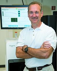
“Until now, we’ve really understood very little about therapy-related AML and why it is so difficult to treat,” said corresponding author Daniel Link, MD, a hematologist/oncologist at Siteman Cancer Center at Washington University and Barnes-Jewish Hospital. “This gives us some important clues for further studies aimed at treatment and prevention.”
The researchers initially sequenced the genomes of 22 cases of therapy-related AML, finding that those patients had similar numbers and types of genetic mutations in their leukemia cells as other patients who developed AML without exposure to chemotherapy or radiation therapy, an indication that cancer treatment does not cause widespread DNA damage.

“This is contrary to what physicians and scientists have long accepted as fact,” said senior author Richard K. Wilson, PhD, the Alan A. and Edith L. Wolff Distinguished Professor of Medicine and director of The Genome Institute at Washington University. “It led us to consider a novel hypothesis: P53 mutations accumulate randomly as part of the aging process and are present in blood stem cells long before a patient is diagnosed with therapy-related AML.”
When therapy-related AML occurs, it typically develops one to five years after treatment with chemotherapy or radiation. Its incidence varies by cancer type. For example, 10 percent of patients with lymphoma that recurs after chemotherapy go on to develop therapy-related AML; among breast cancer patients, only 0.1 percent experience this devastating complication of cancer treatment.
Researchers have known that patients with therapy-related AML are more likely than other AML patients to have a high rate of P53 mutations in their blood cells. The gene is a tumor suppressor and normally works to keep cell division in check and maintain the structure of chromosomes inside cells. But when both copies of the gene are disabled by mutations, cancer can develop.
Surprisingly, when the researchers analyzed blood samples from 19 healthy people ages 68-89 with no history of cancer or chemotherapy, they found that nearly 50 percent had mutations in one copy of P53, an indicator that many people acquire mutations in this gene as they age.
“Most of the time, these mutations are harmless because they only affect one copy of the gene,” Wilson explained.
The finding encouraged the researchers, led by first author Terrence Wong, MD, PhD, a clinical fellow in oncology, to dig further.
Blood stem cells originate in the bone marrow, so the researchers scoured the country to find bone marrow samples from patients with therapy-related AML that had been stored before the patients developed leukemia.
“We wanted to know whether we could go back in time – before a patient is diagnosed with therapy-related AML – to find the exact P53 mutation that caused them to develop leukemia years later,” said Link, the Alan A. and Edith L. Wolff Professor of Medicine.
The researchers found seven bone marrow samples that fit the criteria, and in four of those, they detected specific mutations in P53 that were present at very low rates in blood cells or bone marrow three to six years before the development of leukemia.
In the three cases in which P53 mutations could not be found, the researchers said it’s possible the mutations were present but at rates too low to be detected or that other age-related mutations may have contributed to the onset of therapy-related AML.
In related work in mice, they also showed that chemotherapy causes blood stem cells with mutations in P53 to divide rapidly, which gives them a competitive advantage. But that was not the case in blood stem cells with both copies of the gene intact.
The researchers suspect that the early accumulation of P53 mutations in blood stem cells likely contributes to the frequent chromosomal and genetic abnormalities seen in patients with therapy-related AML and their poor responses to chemotherapy. They suspect that other age-related mutations also may be involved in the disease.
“We’re already conducting follow-up studies to look for other age-related mutations that may be at play in therapy-related AML,” Link said. “As individuals, we’re not genetically homogeneous throughout our lives. Our DNA is constantly changing as we age, and we know this plays an important role in the development of cancer. With advanced genomics, we can investigate the interplay between aging and the random accumulation of mutations, as a means to improve the diagnosis, treatment and prevention of cancer.”
Wong TN, Ramsingh G, Young AL, Miller CA, Touma W, Welch JS, Lamprecht TL, Shen D, Hundal J, Fulton RS, Heath S, Baty JD, Klco JM, Ding L, Mardis ER, Westervelt P, DiPersio JF, Walter MJ, Graubert TA, Ley TJ, Druley T, Link DC and Wilson RK. Role of TP53 mutations in the origin and evolution of therapy-related acute myeloid leukemia. Nature. Dec. 8, 2014.
Washington University School of Medicine’s 2,100 employed and volunteer faculty physicians also are the medical staff of Barnes-Jewish and St. Louis Children’s hospitals. The School of Medicine is one of the leading medical research, teaching and patient-care institutions in the nation, currently ranked sixth in the nation by U.S. News & World Report. Through its affiliations with Barnes-Jewish and St. Louis Children’s hospitals, the School of Medicine is linked to BJC HealthCare.