Scientists are natural problem solvers, full of innovative ideas. But moving those ideas from the laboratory to the marketplace can be difficult, even for those with an entrepreneurial bent.
In part, that’s because federal research dollars typically don’t support the proof-of-concept studies needed to demonstrate the feasibility of a promising new technology or diagnostic test. And while most scientists feel right at home in the laboratory, they often struggle to develop a successful pitch or execute a business plan.
To fill the gap, Washington University’s Bear Cub program provides university scientists with funding to help commercialize their discoveries. Beginning this year, scientists who are funded through the program also have access to business mentors and other hands-on assistance to develop their technologies.
“We want our faculty and students to have every opportunity to commercialize their technologies,” says Bradley Castanho, PhD, director of the university’s Office of Technology Management. “Part of that means creating an atmosphere where scientists are supported and encouraged in their efforts to become entrepreneurs, while also helping to make funding available so they can move their discoveries beyond the lab.”
The university recently announced a new round of Bear Cub funding, with $204,000 going to five scientists:
David Beebe, PhD, the Janet and Bernard Becker Professor of Ophthalmology and Visual Sciences, is developing a way to prevent the formation of cataracts in patients undergoing retinal surgery. To repair the retina, surgeons must remove a portion of the vitreous gel that fills the eye, a process that exposes the lens to oxygen and increases the likelihood of cataracts.
Working with colleagues at Purdue University who developed a novel biological polymer, Beebe will evaluate whether the polymer can preserve the remaining vitreous gel and restore its properties to prevent cataracts from forming. He is now proposing to test the polymer in animal models, with the goal of developing a sterile synthetic polymer powder that could be mixed with sterile saline and infused into the eye at the end of retina surgery. Annually, some 300,000 patients in the U.S. alone could benefit from the technology, the researchers have estimated.
Joseph Gaut, MD, PhD, assistant professor of pathology and immunology, has developed a test for the early detection of acute kidney injury, a complication that can occur in critically ill patients and in those undergoing heart bypass surgery. Some 700,000 U.S. patients undergo heart bypass surgery every year, and one-fourth of them develop kidney damage, which leads to longer hospital stays and deaths, in some cases.
The test developed by Gaut and his colleagues is based on a kidney-specific protein that is elevated in the blood soon after acute kidney damage occurs, typically several days before currently available tests. The researchers will evaluate whether the protein can accurately diagnose early kidney damage in animal models and in heart bypass patients, which would enable earlier treatment.
Michael S. Hughes, PhD, research associate professor of medicine, is working with John E. McCarthy, PhD, the Spencer T. Olin Professor of Mathematics, and Samuel A. Wickline, MD, professor of medicine, to develop an imaging technology that captures certain aspects of electromagnetic and acoustic waves and converts that information into an image.
Rather than being based on wave energy, however, the image measures the entropy, or disorder, in an object and can detect features that are not picked up by ultrasound, CT scans and other conventional imaging. Entropy imaging could potentially have wide applications in medicine and be used to identify defects in materials used by the aerospace and other transportation industries or in heavy manufacturing. Another possible application is in security scanning to detect potential threats and in remote surveillance.
Eric Leuthardt, MD, associate professor of neurological surgery, has designed a monitor to noninvasively detect obstructions in vascular grafts and shunts. The monitor uses a nanoscale flow sensor that can be integrated into an implantable shunt or graft. Both can narrow over time and become obstructed, leading to life-threatening complications.
For example, about one in 500 babies is born with hydrocephalus, a buildup of fluid on the brain. It is most often treated surgically by inserting a shunt that diverts the fluid to another area of the body. But symptoms of pain in the head, even something like a headache, can lead doctors to order CT scans, nuclear medicine studies and sometimes exploratory surgery to determine whether the pain is related to an obstruction.
The sensor Leuthardt has developed can be activated by light to measure the flow rate of fluids through grafts and shunts, and he plans to test the device in animal models.
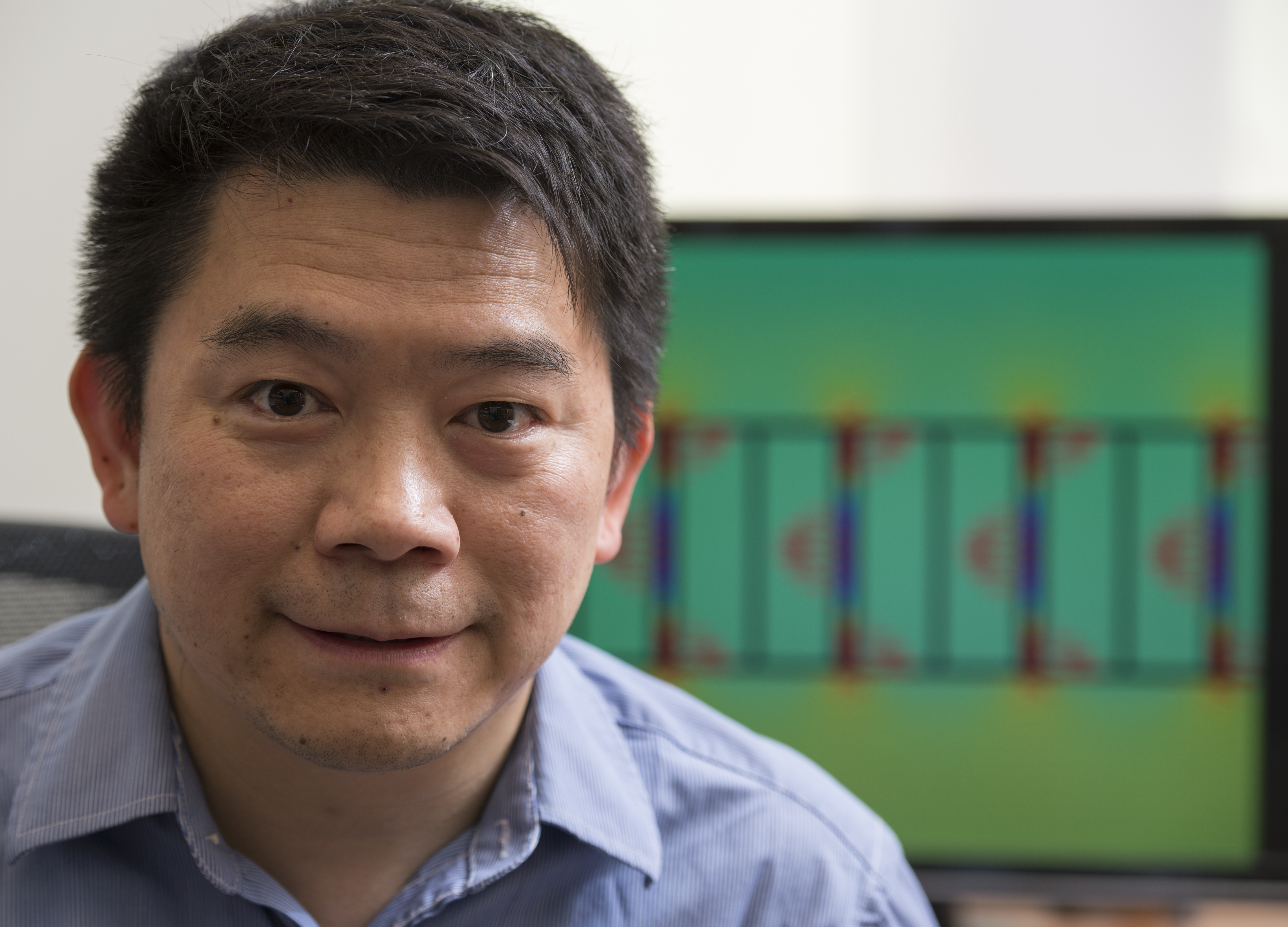
Jung-Tsung Shen, PhD, assistant professor of electrical and systems engineering, has developed a photonic switch that is orders of magnitude faster, smaller and more energy efficient than other switches typically used to support the information superhighway. In the future, demands for broadband signal transmission and processing will require ultra-fast and extremely low-energy optical switching and modulation rates that aren’t possible with current approaches.
The switch designed by Shen and his colleagues uses artificially engineered materials, called metamaterials, that exhibit exceptional optical properties not easily observed in nature. In addition to telecommunications, the switch also could be used in high-resolution medical imaging and in semiconductor manufacturing. Bear Cub funding will allow Shen to further develop and test the switch.