An international team of scientists including B. Duygu Özpolat at Washington University in St. Louis has published the first single-cell atlas for Pristina leidyi (Pristina), the water nymph worm, a segmented annelid with extraordinary regenerative abilities that has fascinated biologists for more than a century.
Annelid worms — including the most familiar among them, the earthworms — are a broadly distributed, highly diverse, economically and environmentally important group of animals.
Most annelids can regenerate missing body parts, and many are able to reproduce asexually. But the adult stem cell populations involved in these processes, as well as the diversity of cell types generated by the stem cells, have remained unknown.
This particular worm, Pristina, first caught the eye of biologists in the 1800s and has remained an object of much interest. Under laboratory conditions, Pristina grows very rapidly and creates copies of itself by asexual reproduction.

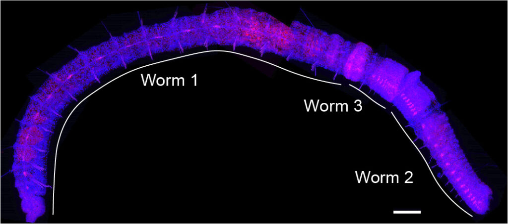
Using a mechanism called paratomic fission, the worm starts forming and differentiating new head and tail segments from within a single body segment, producing what is known as a “chain of worms.” Eventually these clones separate and become distinct individuals.
“These worms are constantly generating all body parts and therefore all adult cell types,” said Özpolat, an assistant professor of biology in Arts & Sciences.

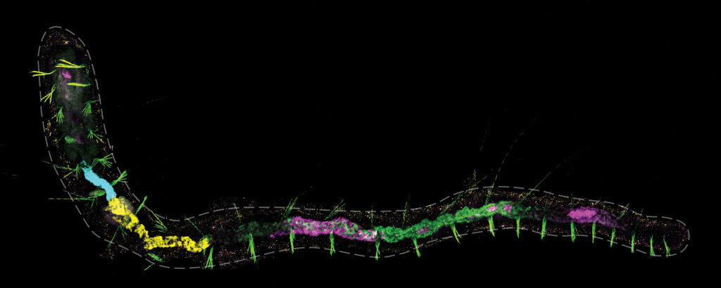
In all, the new single-cell atlas for this worm assembles 75,218 single-cell transcriptomes characterizing all major annelid cell types, including complex patterns of regionally expressed genes in the annelid gut, as well as neuronal, muscle and epidermal specific genes. The study is published in Nature Communications.
“We want to understand how different organisms like Pristina have evolved to continuously grow throughout their lives and regenerate, the nature of cells involved in these processes, and molecular signatures they have,” Özpolat said.
“Different organisms have evolved different strategies,” she said. “The cellular and genetic mechanisms we learn from the worms not only help us understand these fascinating organisms better, but can also inform stem cell technologies and regenerative medicine down the line.”
“It is curious that these worms can maintain adult stem cells indefinitely,” Özpolat said.
“We have grown thousands of clones from a single individual, and our worm cultures are still going strong.”
Segmented reality
Özpolat produced and mapped the single-cell atlas for Pristina in partnership with Jordi Solana at University of Exeter in the United Kingdom and Patricia Álvarez-Campos at Universidad Autónoma de Madrid. Solana and his group had previously focused on stem cells in a different type of worm: the planarian, or flatworm.
Pristina was a new challenge for the combined team to take on. Annelids like Pristina have bodies that are made up of a series of segments with a growth zone at the tail end which produces new segments continuously from two concentric rings of stem cells.
And then there was Pristina’s unusual tendency to bud, or make a chain.
“When the animal reaches a certain size, then it somehow senses that it has reached the threshold to split,” Özpolat said. “And so, it starts making a head and then a new tail in the middle of its body. This means it has to completely reorganize what used to be a segment that contained the intestine into a segment that now will have a new brain, or new ovaries and testes.”
As a postdoctoral scholar, Özpolat was most interested in how worms made new gonads as part of this chain-like reproductive process.
For her future research directions, she plans to also focus on the gut.

“With single-cell atlases, you take an entire organism and you literally split it into its individual cells. And then you look at gene expression in each cell separately,” she said. “Then you group them into these maps, based on their similarities in terms of gene expression patterns.
“We found cell types that we didn’t even know that existed in this animal,” Özpolat said. “Its gut is so neatly organized and specific. There are about 12 different gut cell types in this tiny little worm, which will be very interesting for future projects we’re already working on.
“It just opens up so many doors now that you can visualize these different cell types, and how they behave during fission and regeneration,” she said.


