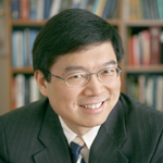
Lihong Wang, PhD, has received a National Institutes of Health (NIH) Director’s Pioneer Award to explore novel imaging techniques using light that promise significant improvements in biomedical imaging and light therapy.

One of only 11 recipients of the highly competitive award, Wang was selected from among 600 applicants. The award supports individual scientists of exceptional creativity who propose pioneering — and possibly transforming — approaches to major challenges in biomedical and behavioral research, according to the NIH.
The award will provide Wang with a total budget of $3.8 million over five years.
Wang, the Gene K. Beare Distinguished Professor of Biomedical Engineering at Washington University in St. Louis, says his research will explore transporting light into the body’s tissues far beyond the classical penetration limits for high-sensitivity imaging and low-side-effect therapy.
“I am honored to have received this award from among such a competitive group,” Wang says. “This award will allow us the intellectual freedom and resources to develop a brand new technology. If successfully implemented, it would impact many disciplines of biomedicine with applications, including imaging, such as functional brain imaging and reporter gene imaging; sensing (oximetry and glucometry); manipulation (optogenetics and nerve stimulation); and therapy (photodynamic therapy and photothermal therapy).”
A leading researcher on new methods of cancer imaging, Wang has received more than 30 research grants as the principal investigator with a cumulative budget of more than $38 million. His research on non-ionizing biophotonic imaging has been supported by the NIH, National Science Foundation (NSF), the U.S. Department of Defense, The Whitaker Foundation and the National Institute of Standards and Technology.
“I am extremely pleased and proud that Lihong is receiving this award which recognizes his tremendous creativity and innovativeness,” says Frank Yin, MD, PhD, the Stephen F. and Camilla T. Brauer Distinguished Professor of Biomedical Engineering and chair of the department.
“His pioneering work in developing photoacoustic tomography, along with its many variations and combinations, has spawned a whole new field. The ability to safely image deep into the body with extraordinarily high spatial and temporal resolution — at a fraction of the cost of conventional imaging methods — promises to revolutionize medical imaging in the years to come. I look forward to seeing the many new developments that will arise from his work.”
Wang and his lab founded a type of medical imaging that gives physicians a new look at the body’s internal organs, publishing the first paper on the technique in 2003.
Called photoacoustic tomography, the technique relies on light and sound to create detailed, color pictures of tumors deep inside the body and may eventually help doctors diagnose cancer earlier than is now possible and to more precisely monitor the effects of cancer treatment — all without the radiation involved in X-rays and CT scans or the expense of MRIs.
Wang, who is affiliated with the Alvin J. Siteman Cancer Center at Barnes-Jewish Hospital and Washington University School of Medicine, is working with Washington University physicians to evaluate the technology for four uses: identifying the sentinel lymph nodes for breast cancer staging, which may eliminate the need for surgical lymph node biopsies; monitoring early response to chemotherapy; imaging melanomas; and imaging the gastrointestinal tract.
Wang also invented a “guide star” for biomedical imaging that allows scientists to focus light to a controllable position within the body’s tissue. Wang’s guide star is an ultrasound beam that “tags” light that passes through it.
The technique, called time-reversed ultrasonically encoded (TRUE) optical focusing, allows scientists to focus light to a controllable position within tissue. Wang says TRUE will lead to more effective light imaging, sensing, manipulation and therapy, all of which could benefit medical research, diagnostics and therapeutics.
He has published more than 300 peer-reviewed articles and is a frequent keynote speaker at major conferences and symposia. His Monte Carlo model of photon transport in scattering media has been used worldwide. He serves as the editor-in-chief of a premier journal in biomedical optics. He wrote one of the first textbooks in his field, which won the Joseph W. Goodman Book Writing Award.
He received the NIH FIRST and the NSF’s CAREER Award. He also received The Optical Society’s C. E. K. Mees Medal and IEEE’s Technical Achievement Award for seminal contributions to photoacoustic tomography and Monte Carlo modeling of photon transport in biological tissues and for leadership in the international biophotonics community.