Five minutes in a scanner can reveal how far a child’s brain has come along the path from childhood to maturity and potentially shed light on a range of psychological and developmental disorders, scientists at Washington University School of Medicine in St. Louis have shown.
Researchers assert this week in Science that their study proves brain imaging data can offer more extensive help in tracking aberrant brain development.
“Pediatricians regularly plot where their patients are in terms of height, weight and other measures, and then match these up to standardized curves that track typical developmental pathways,” says senior author Bradley Schlaggar, MD, PhD, a Washington University pediatric neurologist. “When the patient deviates too strongly from the standardized ranges or veers suddenly from one developmental path to another, the physician knows there’s a need to start asking why.”
Schlaggar and his colleagues say a new way of looking at brain scanning data may be able to provide similar guidance for monitoring and treating of patients with psychiatric and developmental disorders.
Schlaggar, the A. Ernest and Jane G. Stein Associate Professor of Neurology, says he has sent children with obvious, profound psychiatric conditions for MRI scans and received results marked “no abnormalities noted.”
“That’s typically looking at the data from a structural point of view—what’s different about the shapes of various brain regions,” he says. “But MRI also offers ways to analyze how different parts of the brain work together functionally.”
Compare functional data to standardized models of how brain function or disease normally develops, Schlaggar says, and a range of new clinical insights becomes available.
Schlaggar and his colleagues use an approach to brain scanning called resting state functional connectivity. By correlating increases and decreases in blood flow to the various brain regions as subjects rest in the scanner, scientists determine which of these regions work together in brain networks.
In a study published in 2009, Washington University scientists showed that as the brain matures, these brain networks change (see http://news.wustl.edu/news/Pages/14199.aspx). The overall organization switches from networks involving regions physically close to each other, which is the dominant motif in a child’s brain, to networks that connect distant regions, the primary organizational principal in adult brains.
For the new study, lead author Nico Dosenbach, MD, PhD, a pediatric neurology resident at St. Louis Children’s Hospital, took this and other distinctions that mark the transition from child to adult brain and adapted them for use in a technique for mathematical analysis called a support vector machine. The technique is employed in many contexts in science and economics and on the Internet.
“It’s a way that mathematicians have developed for predicting something with high specificity and sensitivity when you have huge amounts of data instead of one really good measurement,” Dosenbach explains. “Any one of these measurements doesn’t tell you much, but if you put them together and use the right math to sift through and restructure them, you can get good predictive results.”

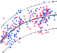
Dosenbach used data from five-minute MRI scans of 238 normal subjects ranging in age from 7 to 30. The support vector machine analyzed approximately 13,000 functional brain connections and selected the best 200 to produce a single index of the maturity of each subject. The data allowed scientists to predict whether subjects were children or adults, and roughly formed a curving line that tracks the path of normal functional brain development.
The researchers suspect patients with brain disorders will appear out of alignment with this normal developmental curve.
“The beauty of this approach is that it lets you ask what’s different in the way that children with autism, for example, are off the normal development curve versus the way children with attention-deficit disorder are off that curve,” Schlaggar says.
Schlaggar suggests that functional brain scans might be conducted on a group of children at risk but not yet suffering from a developmental disorder.
“When a fraction of them later develop that disorder, you can go back and construct an analysis like this one that will help predict the characteristics of the next child at highest risk of developing the disorder,” he says. “That’s very powerful both clinically and from the perspective of understanding the causes of these disorders.”
This approach might enable treatment prior to onset of symptoms, Schlaggar says, and should help physicians more quickly and closely track the results of clinical trials of new therapies.
“MRI scans are expensive, so this may not be what we use for everyone right now,” Dosenbach says. “But many children with these types of disorders already receive regular structural MRI scans, and five more minutes in the scanner won’t add that much to the cost.”
Dosenbach NUF, Nardos B, Cohen AL, Fair DA, Power JD, Church JA, Nelson SM, Wig GS, Vogel AC, Lessov-Schlaggar CN, Barnes KA, Dubis JW, Feczko E, Coalson RS, Pruett JR JR, Barch DM, Petersen SE, Schlaggar BL. Prediction of individual brain maturity using fMRI. Science, Sept. 10, 2010.
Funding from the National Institutes of Health, the John Merck Scholars Fund, the Burroughs-Wellcome Fund, the Dana Foundation, the Ogle Family Fund, the McDonnell Center, the Simons Foundation, the American Hearing Research Foundation, and the Diabetes Research Center at Washington University supported this research.
Washington University School of Medicine’s 2,100 employed and volunteer faculty physicians also are the medical staff of Barnes-Jewish and St. Louis Children’s hospitals. The School of Medicine is one of the leading medical research, teaching and patient care institutions in the nation, currently ranked fourth in the nation by U.S. News & World Report. Through its affiliations with Barnes-Jewish and St. Louis Children’s hospitals, the School of Medicine is linked to BJC HealthCare.