Investigators at Washington University School of Medicine in St. Louis have identified a genetic variation associated with an earlier age of onset in Alzheimer’s disease.

Unlike genetic mutations previously linked to rare, inherited forms of early-onset Alzheimer’s disease — which can strike people as young as their 30s or 40s — these variants influence an earlier presentation of symptoms in people affected by the more common, late-onset form of the disease.
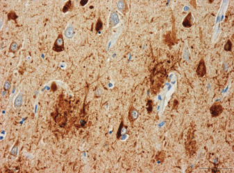
Two principal features characterize Alzheimer’s disease in the brain: amyloid plaques and neurofibrillary tangles. The plaques contain a protein called amyloid-beta. The tangles are made of a protein called tau.
The research team analyzed DNA from 313 subjects from Washington University’s Alzheimer’s Disease Research Center (ADRC), focusing on locations in the tau gene that previously have been found to vary between people.

“We focused on this gene for two reasons: First, it codes for the tau protein that we find in neurofibrillary tangles, and secondly, some studies in the scientific literature show an association between the gene and Alzheimer’s disease, while others do not,” says principal investigator Alison M. Goate, D. Phil., the Samuel and Mae S. Ludwig Professor of Genetics in Psychiatry and professor of neurology. “Even a study from our own group had found no association between tau gene variants and Alzheimer’s disease.”
But this study, reported in the June 10 issue of the Proceedings of the National Academy of Sciences, changes that. One reason past studies may have produced conflicting results is that most, if not all, people have amyloid plaques in the brain years before they develop clinical symptoms of Alzheimer’s.
“It’s not uncommon for us to determine that an older person is fully intact mentally only to find the presence of substantial Alzheimer’s pathology on examining that person’s brain after death,” says John C. Morris, M.D., the Harvey A. and Dorismae Friedman Distinguished Professor of Neurology and director of the ADRC and of the Harvey A. Friedman Center for Aging. “We suspect that Alzheimer lesions may be present in the brain long before we can detect any clinical symptoms.”
Previous research from Goate’s colleagues David M. Holtzman, M.D., the Andrew B. and Gretchen P. Jones Professor and head of the Department of Neurology, and Anne M. Fagan, Ph.D., associate professor of neurology, measured soluble forms of amyloid-beta and tau proteins in the cerebrospinal fluid. They determined that amyloid-beta levels indicate whether or not amyloid plaques are present in the brain.
“A particular form of amyloid-beta called amyloid-beta 42, tends to be higher in the cerebrospinal fluid of normal individuals and lower in patients with Alzheimer’s disease and in cognitively normal people who have amyloid plaques in the brain,” says Holtzman. “Tau protein levels in the cerebrospinal fluid increase when a person starts developing dementia.”
Finding those amyloid deposits once required examination of the brain after a person’s death, but researchers now can detect their presence by assessing them with positron emission tomography (PET) imaging as well as measuring amyloid beta 42. When PET imaging detects amyloid in the brain, patients have lower levels of amyloid-beta 42 in their cerebrospinal fluid.
Goate’s team found that four DNA sequence variants in the tau gene were associated with higher levels of tau protein in the cerebrospinal fluid. Then they divided patients into two groups. One group had evidence of plaques in the brain, while the other did not. The investigators found that the variations in the gene are only associated with an increase in tau protein levels in the cerebrospinal fluid when there is evidence of amyloid plaques in the brain.
Armed with those findings, Goate’s team predicted that the variants in the tau gene that contributed to higher levels of tau protein in the cerebrospinal fluid would be associated with a younger age at the onset of Alzheimer’s disease symptoms.
“So we went back to the ADRC’s clinical samples, and that’s exactly what we found,” she says. “Individuals who carry these genetic variations that lead to higher levels of tau in cerebrospinal fluid actually have an earlier age of onset than those who carry variants that are associated with lower levels of tau.”
Goate says these sequence variants in the tau gene are not linked to risk of Alzheimer’s disease but rather to earlier cognitive problems once plaques have started to form in the brain. She says people who possess those genetic variants, if they are fated to develop Alzheimer’s disease, will experience symptoms sooner.
“Advanced techniques in identifying markers for amyloid and tau deposition in the brains of people with Alzheimer’s disease, in combination with genetic analysis, are giving us new clues about how the disease begins,” says Marcelle Morrison-Bogorad, Ph.D., director of the Division of Neuroscience at the National Institute on Aging. “This study offers important information about the levels of expression of particular forms of the tau gene in the presence of brain amyloid and therefore may help us understand why the disease begins earlier in some persons than in others.”
Goate says these findings lend further support to the hypothesis that amyloid-beta plaques form earlier in the cascade of Alzheimer’s pathology, and that the tau protein is involved in how the disease progresses. She says more work is needed at the cellular level to figure out how the proteins interact to cause Alzheimer’s symptoms, but in the meantime, she says identifying these variants in the tau gene may provide clinicians with a new target for potential therapies.
“Even when there already is evidence of amyloid deposition in the brain, if we could find a way to lower tau levels, we would predict that the onset of symptoms may be delayed,” she says. “But we need to do a lot more cell biology and research in animal models before we can hope to do that.”
Kauwe JSK, Cruchaga C. Mayo K, Fenoglio C, Bertelsen S, Nowotny P, Galimberti D, Scarpini E, Morris JC, Fagan AM, Holtzman DM, Goate AM. Variation in MAPT is associated with cerebrospinal fluid tau levels in the presence of amyloid-beta deposition. Proceedings of the National Academy of Sciences, vol. 105 (23). pp. 8050-8054. June 10, 2008 www.pnas.org/cgi/doi/10/1073/pnas.0801227105
This study was supported by the National Institute on Aging of the National Institutes of Health, the Barnes-Jewish Foundation, the Associazione “Amici Del Centro Dino Ferrari,” Monzino Foundation, Ing. Cesare Cusan and a Ford Foundation Fellowship.
Washington University School of Medicine’s 2,100 employed and volunteer faculty physicians also are the medical staff of Barnes-Jewish and St. Louis Children’s Hospitals. The School of Medicine is one of the leading medical research, teaching, and patient care institutions in the nation, currently ranked third in the nation by U.S. News & World Report. Through its affiliations with Barnes-Jewish and St. Louis Children’s Hospitals, the School of Medicine is linked to BJC HealthCare.