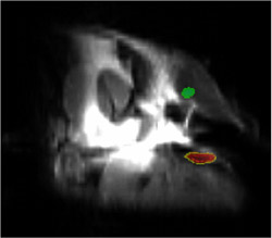
To the delight of researchers at Washington University School of Medicine in St. Louis, living cells gobbled up fluorine-laced nanoparticles without needing any coaxing. Then, because of the unusual meal, the cells were easily located with MRI scanning after being injected into mice.

Developed in the laboratories of Samuel A. Wickline, M.D., and Gregory Lanza, M.D., Ph.D., the nanoparticles could soon allow researchers and physicians to directly track cells used in medical treatments using unique signatures from the ingested nanoparticle beacons.
In an article that will appear in the June issue of the FASEB Journal, lead author Kathryn C. Partlow, a doctoral student in Wickline’s lab, describes using perfluorocarbon nanoparticles to label endothelial progenitor cells taken from human umbilical cord blood. Such cells can be primed to help build new blood vessels when injected into the body. The researchers believe nanoparticle-labeled stem cells like these could prove useful for monitoring tumors and diagnosing and treating cardiovascular problems.
The nanoparticles contain a fluorine-based compound that can be detected by MRI scanners. Fluorine is most commonly known for being an element included in fluoride toothpastes. Wickline, who heads the Siteman Center of Cancer Nanotechnology Excellence, says this technology offers significant advantages over other cell-labeling technologies under development.
“We can tune an MRI scanner to the specific frequency of the fluorine compound in the nanoparticles, and only the nanoparticle-containing cells will be visible in the scan,” he says. “That eliminates any background signal, which often interferes with medical imaging. Moreover, the lack of interference means we can measure very low amounts of the labeled cells and closely estimate their number by the brightness of the image.”
The researchers believe that nanoparticle-labeled adult stem cells could be used to evaluate tumors. Under an MRI scan, the presence of the labeled cells would reveal that the tumor was adding new blood vessels and therefore aggressively growing.
Adult stem cells are also under investigation in therapies that enhance new blood vessel growth to improve the blood supply to diabetic patients’ limbs or to repair blood vessels after a heart attack or bypass surgery. Tracking nanoparticle-labeled cells used in such treatments by MRI imaging would allow physicians to monitor the treatment’s success or failure.
The nanoparticles — called “nano” because they measure only about 200 nanometers across, or 500 times smaller than the width of a human hair — are made up largely of perfluorocarbon, a safe compound used in artificial blood. The fluorine atoms in the particles can be detected by tuning an MRI scanner to the unique signal frequency emitted by the perfluorocarbon compound used.
Since several perfluorocarbon compounds are available, different types of cells potentially could be labeled with different compounds, injected and then detected separately by tuning the MRI scanner to each one’s individual frequency, says Wickline.
That makes the labeled cells potentially useful for vascular research as well. “Many kinds of cells are involved in the formation of new blood vessels,” Partlow says. “Because we can create a separate MRI signature for different cells with these various types of unique nanoparticles, we could use them to better understand each cell type’s role.”
The nanoparticles are very compatible with living cells, according to the research findings. “The cells just take these particles in naturally — no special sauces have to be added to make them tasty to these cells,” says Wickline, also professor of medicine, of physics and of biomedical engineering and a Washington University heart specialist at Barnes-Jewish Hospital. “And then the cells just go about their business and do what they’re supposed to do by homing in on targeted regions of the body.”
Laboratory tests showed that the cells retained their usual surface markers and that they were still functional after the labeling process. The labeled cells were shown to migrate to and incorporate into blood vessels forming around tumors in mice.
The researchers believe the cells could soon be used in clinical settings. “Kathy and colleagues showed that we can scan for these cells at the same MRI field strength we are using in medical imaging,” Wickline says. “Although we reported the first use of perfluorocarbon molecular imaging for detection of certain pathologies a few years ago, no one would have predicted that you could get enough signal from such small quantities of perfluorocarbons in labeled stem cells to actually see them. I think we’ve dispelled that notion, and the fluorine imaging approach already is becoming more popular for molecular imaging of various cell and tissue types.”
Next the research group will evaluate how nanoparticle-labeled cells function in living organisms. “We’ll track injected cells in real time and see where they accumulate and how long they live,” Partlow says. “Then we’ll go on to investigate how they work in therapeutic applications.”
Partlow KC, Chen J, Brant JA, Neubauer AM, Meyerrose TE, Creer MH, Nolta JA, Caruthers SD, Lanza GM, Wickline SA. 19F magnetic resonance imaging for stem/progenitor cell tracking with multiple unique perfluorocarbon nanobeacons. FASEB J 2007 Feb 6 (advanced online publication).
Funding from the National Institutes of Health and the American Heart Association supported this research.
Washington University School of Medicine’s full-time and volunteer faculty physicians also are the medical staff of Barnes-Jewish and St. Louis Children’s hospitals. The School of Medicine is one of the leading medical research, teaching and patient care institutions in the nation, currently ranked fourth in the nation by U.S. News & World Report. Through its affiliations with Barnes-Jewish and St. Louis Children’s hospitals, the School of Medicine is linked to BJC HealthCare.
Siteman Cancer Center is the only NCI-designated Comprehensive Cancer Center within a 240-mile radius of St. Louis. Siteman Cancer Center is composed of the combined cancer research and treatment programs of Barnes-Jewish Hospital and Washington University School of Medicine.