Lew and his students build advanced imaging systems to study biological and chemical systems at the nanoscale, leveraging innovations in applied optics, signal and image processing, design optimization, and physical chemistry.
Their advanced nanoscopes (microscopes with nanometer resolution) visualize the activity of individual molecular machines inside and outside living cells. Examples of new technologies developed in the Lew Lab include 1) using tiny fluorescent molecules as sensors that can detect amyloid proteins, 2) designing new “lenses” to create imaging systems that can visualize how molecules move and tumble, and 3) new imaging software that minimizes artifacts in super-resolution images.
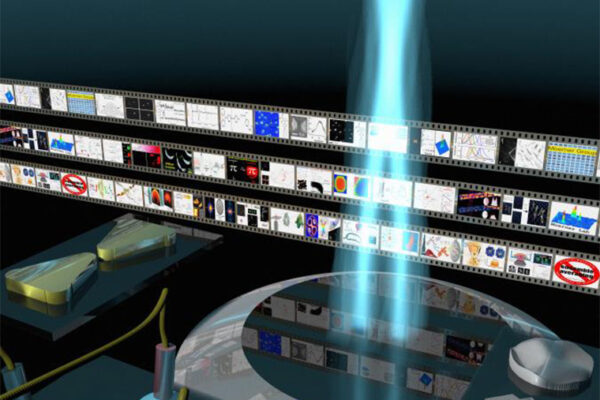
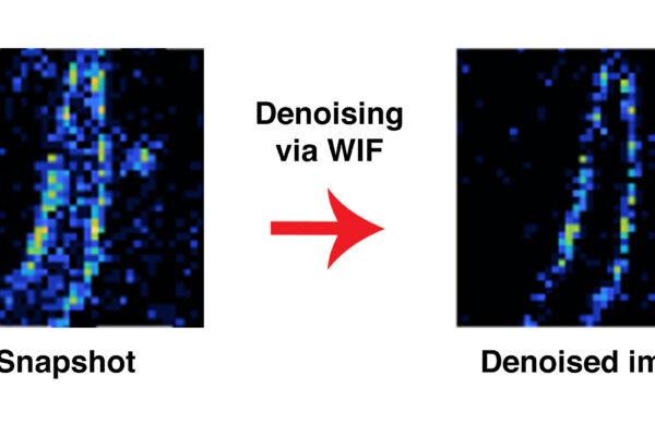
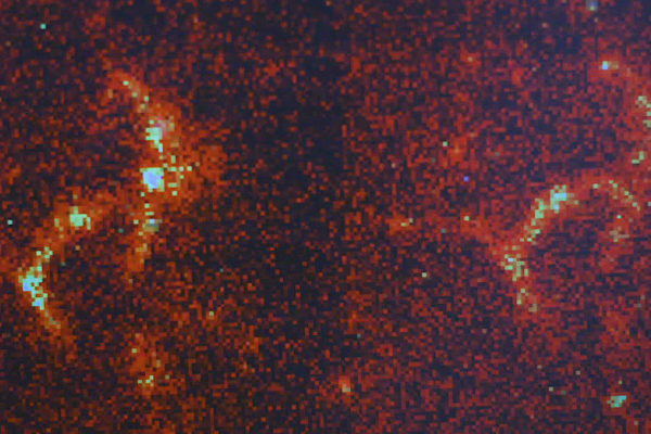
Using machine learning with an additional processing step, researchers from the lab of Matthew Lew at the McKelvey School of Engineering can wrest a host of information from a few pixels of light.
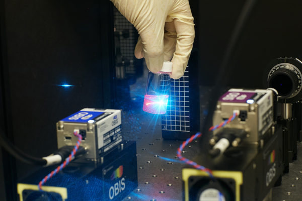
Researchers in the Lew lab in the McKelvey School of Engineering are using light in novel ways to better image biological samples.
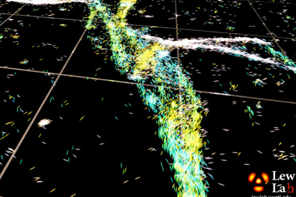
A new imaging technology from the lab of Matthew Lew at the McKelvey School of Engineering uses polarized “optical vortices” to provide a detailed, dynamic view of molecules in motion.
A new computational method from the McKelvey School of Engineering helps scientists validate the accuracy of microscopic images.
Using properties of light from fluorescent probes is at the heart of a new imaging technique developed at Washington University’s McKelvey School of Engineering that allows for an unprecedented look inside cell membranes.
A new technique developed in the lab of Matthew Lew at the McKelvey School of Engineering at Washington University in St. Louis measures the orientation of single molecules. It is enabling, for the first time, optical microscopy to reveal nanoscale details about the structures of these problematic proteins.
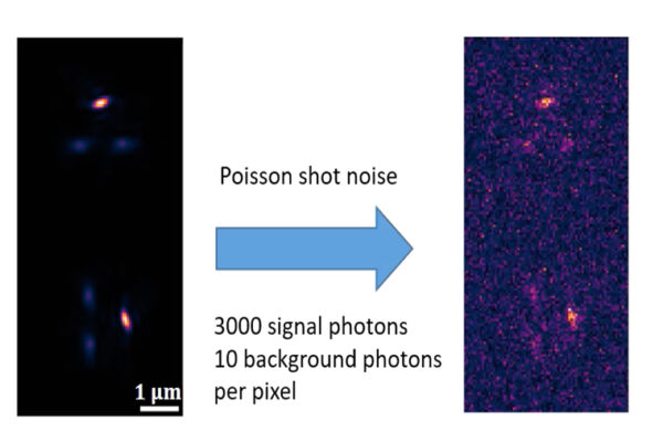
As researchers probe smaller parts of our world, a “picture” is not always showing what it may seem to show. One researcher at the McKelvey School of Engineering at Washington University in St. Louis has uncovered a fundamental limit to our ability to trust what we see when it comes to images of molecular motion.
Tiny protein structures called amyloids are key to understanding certain devastating age-related diseases, but they are so minuscule they can’t be seen using conventional microscopic methods. A team of engineers at Washington University in St. Louis has developed a new technique that uses temporary fluorescence, causing the amyloids to flash or “blink”, allowing researchers to better spot these problematic proteins.
An engineer at Washington University in St. Louis plans to push the envelope of microscopic imaging, to better visualize the molecules involved in Alzheimer’s disease.


