A faculty member at Washington University in St. Louis’ School of Engineering & Applied Science has been awarded two separate grants worth a combined $2.5 million to develop better biomedical imaging tools.

Mark Anastasio, professor of biomedical engineering, will use a four-year, $2.2 million grant from the National Institutes of Health (NIH) to create a new X-ray technique that will assist engineers as they develop new bioengineered tissues.
“We are developing a new imaging technology based on phase-contrast X-ray imaging,” Anastasio said. “It will serve as an enabling technology for tissue engineering studies.”
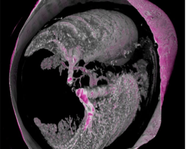
A typical X-ray image forms as radiation that is absorbed by tissues and bones, providing doctors with a look inside the body. Anastasio’s new technology doesn’t rely entirely on the absorption of X-ray energy, it also exploits wave optic effects, measuring the X-ray’s refractions for a much more precise peek inside.
“In some cases, you can make the X-ray beam act like a wave,” Anastasio said. “In such cases, when it hits an interface between two tissues, it can actually bend by a very small angle; it can refract. If you can measure the angle by which these rays refract, you can form a more detailed image based on that information. It will let you see things that would normally be invisible to conventional X-ray imaging.”
That sharper image is especially important when it comes to monitoring bioengineered materials being tested inside small animals. They are made up of mostly water, and difficult to see on a normal X-ray image. This new technique will help bioengineers better view and monitor implanted biomaterials in vivo, which can provide valuable information to help refine tissue engineering strategies.
The NIH grant also will fund development of algorithms and resulting software for the new X-ray technology. Eric Brey, professor of biomedical engineering at the Illinois Institute of Technology, is Anastasio’s partner on the project and will utilize the developed technologies to address important problems in the field of tissue engineering.
A second grant, $345,000 from the National Science Foundation, will fund research on the mathematical side of medical imaging. Anastasio and Jianliang Qian, professor of mathematics and director of the Michigan Center for Industrial and Applied Mathematics at Michigan State University, will develop new mathematical and computational methods for producing 3-D images in photoacoustic and ultrasound tomography.
