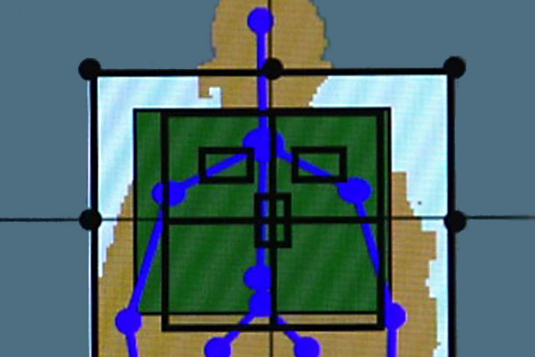
With the aim of producing high-quality X-rays with minimal radiation exposure, particularly in children, researchers have developed a new approach to imaging patients. Surprisingly, the new technology isn’t a high-tech, high-dollar piece of machinery. Rather, it’s based on the Xbox gaming system.
Using proprietary software developed for the Microsoft Kinect system, researchers at Washington University School of Medicine in St. Louis have adapted hands-free technology used for the popular Xbox system to aid radiographers when taking X-rays.
The software coupled with the Kinect system can measure thickness of body parts and check for motion, positioning and the X-ray field of view immediately before imaging, said Steven Don, MD, associate professor of radiology at the university’s Mallinckrodt Institute of Radiology. Real-time monitoring alerts technologists to factors that could compromise image quality. For example, “movement during an X-ray requires retakes, thereby increasing radiation exposure,” Don said.
A feasibility study will be presented Wednesday, Dec. 2, at the Radiological Society of North America’s annual meeting in Chicago.
“The goal is to produce high-quality X-ray images at a low radiation dose without repeating images,” Don said. “It sounds surprising to say that the Xbox gaming system could help us to improve medical imaging, but our study suggests that this is possible.”

The technology could benefit all patients but particularly children because of their sensitivity to radiation and greater variation in body sizes, which can range from premature infants to adult-sized teenagers. Setting appropriate X-ray techniques to minimize radiation exposure depends on the thickness of the body part being imaged. High-quality X-rays are critical in determining diagnoses and treatment plans.
Traditionally steel calipers have been used to measure body-part thickness for X-rays. However, calipers are “time-consuming, intrusive and often scary to kids, especially those who are sick or injured,” said Don, a pediatric radiologist who treats patients at St. Louis Children’s Hospital.
“To achieve the best image quality while minimizing radiation exposure, X-ray technique needs to be based on body-part thickness,” Don said. The gaming software has an infrared sensor to measure body-part thickness automatically without patient contact.
“Additionally, we use the optical camera to confirm the patient is properly positioned,” he explained.
Originally developed as a motion sensor and voice and facial recognition device for the Xbox gaming system, Microsoft Kinect software allows individuals to play games hands-free, or without a standard controller. Scientists, computer specialists and other inventors have since adapted the Xbox technology for nongaming applications.
Don and his colleagues, for example, combined the Microsoft Kinect 1.0 technology with proprietary software to improve X-ray imaging. With help from Washington University’s Office of Technology Management, the team applied for a patent last year.
Don developed the technology with William Clayton, a former computer programmer at the School of Medicine, and Robert MacDougall, a clinical medical physicist at Boston Children’s Hospital.
This year, Don and his colleagues have received funding from Washington University and The Society for Pediatric Radiology. They will use these resources to continue research with the updated Microsoft Kinect 2.0 and seek feedback from radiological technologists to improve the software.
While further research and development are needed, the eventual goal is to apply the technology to new X-ray machines as well as retrofitting older equipment.
“Patients, technologists and radiologists want the best quality X-rays at the lowest dose possible without repeating images,” Don said. “This technology is a tool to help achieve that goal.”
