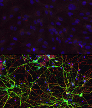
Scientists have described a way to convert human skin cells directly into a specific type of brain cell affected by Huntington’s disease, an ultimately fatal neurodegenerative disorder. Unlike other techniques that turn one cell type into another, this new process does not pass through a stem cell phase, avoiding the production of multiple cell types, the study’s authors report.
The researchers, at Washington University School of Medicine in St. Louis, demonstrated that these converted cells survived at least six months after injection into the brains of mice and behaved similarly to native cells in the brain.
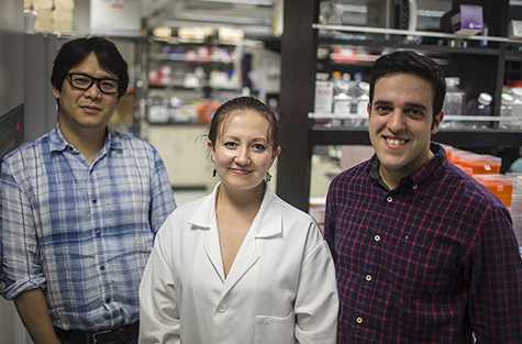
“Not only did these transplanted cells survive in the mouse brain, they showed functional properties similar to those of native cells,” said senior author Andrew S. Yoo, PhD, assistant professor of developmental biology. “These cells are known to extend projections into certain brain regions. And we found the human transplanted cells also connected to these distant targets in the mouse brain. That’s a landmark point about this paper.”
The work appears Oct. 22 in the journal Neuron.
The investigators produced a specific type of brain cell called medium spiny neurons, which are important for controlling movement. They are the primary cells affected in Huntington’s disease, an inherited genetic disorder that causes involuntary muscle movements and cognitive decline usually beginning in middle-adulthood. Patients with the condition live about 20 years following the onset of symptoms, which steadily worsen over time.
The research involved adult human skin cells, rather than more commonly studied mouse cells or even human cells at an earlier stage of development. In regard to potential future therapies, the ability to convert adult human cells presents the possibility of using a patient’s own skin cells, which are easily accessible and won’t be rejected by the immune system.
To reprogram these cells, Yoo and his colleagues put the skin cells in an environment that closely mimics the environment of brain cells. They knew from past work that exposure to two small molecules of RNA, a close chemical cousin of DNA, could turn skin cells into a mix of different types of neurons.
In a skin cell, the DNA instructions for how to be a brain cell, or any other type of cell, is neatly packed away, unused. In past research published in Nature, Yoo and his colleagues showed that exposure to two microRNAs called miR-9 and miR-124 altered the machinery that governs packaging of DNA. Though the investigators still are unraveling the details of this complex process, these microRNAs appear to be opening up the tightly packaged sections of DNA important for brain cells, allowing expression of genes governing development and function of neurons.
Knowing exposure to these microRNAs alone could change skin cells into a mix of neurons, the researchers then started to fine tune the chemical signals, exposing the cells to additional molecules called transcription factors that they knew were present in the part of the brain where medium spiny neurons are common.

“We think that the microRNAs are really doing the heavy lifting,” said co-first author Matheus B. Victor, a graduate student in neuroscience. “They are priming the skin cells to become neurons. The transcription factors we add then guide the skin cells to become a specific subtype, in this case medium spiny neurons. We think we could produce different types of neurons by switching out different transcription factors.”
Yoo also explained that the microRNAs, but not the transcription factors, are important components for the general reprogramming of human skin cells directly to neurons. His team, including co-first author Michelle C. Richner, senior research technician, showed that when the skin cells were exposed to the transcription factors alone, without the microRNAs, the conversion into neurons wasn’t successful.
The researchers performed extensive tests to demonstrate that these newly converted brain cells did indeed look and behave like native medium spiny neurons. The converted cells expressed genes specific to native human medium spiny neurons and did not express genes for other types of neurons. When transplanted into the mouse brain, the converted cells showed morphological and functional properties similar to native neurons.
To study the cellular properties associated with the disease, the investigators now are taking skin cells from patients with Huntington’s disease and reprogramming them into medium spiny neurons using the approach described in the new paper. They also plan to inject healthy reprogrammed human cells into mice with a model of Huntington’s disease to see if this has any effect on the symptoms.
This work was supported by the National Science Foundation Graduate Research Fellowship (DGE-1143954), the fellowship from Cognitive, Computational and Systems Neuroscience Pathway (T32N5023547), and grants from the National Institutes of Health (NIH) (MH078823), including from the National Institute of General Medical Sciences (R01 GM104991) and the National Heart Lung and Blood Institute (T32 HL007275), the NIH Director’s Innovator Award (DP2) and awards from the Mallinckrodt Jr. Foundation, Ellison Medical Foundation, and Presidential Early Career Award for Scientists and Engineers.
Victor MB, Richner M, Hermanstyne TO, Ransdell JL, Sobieski C, Deng PY, Klyachko VA, Nerbonne JM, Yoo AS. Generation of human striatal neurons by microRNA-dependent direct conversion of fibroblasts. Neuron. Oct. 22, 2014.
Washington University School of Medicine’s 2,100 employed and volunteer faculty physicians also are the medical staff of Barnes-Jewish and St. Louis Children’s hospitals. The School of Medicine is one of the leading medical research, teaching and patient-care institutions in the nation, currently ranked sixth in the nation by U.S. News & World Report. Through its affiliations with Barnes-Jewish and St. Louis Children’s hospitals, the School of Medicine is linked to BJC HealthCare.