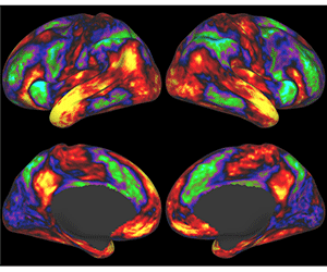
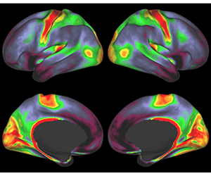
The Human Connectome Project, a five-year endeavor to link brain connectivity to human behavior, has released a set of high-quality imaging and behavioral data to the scientific community. The project has two major goals: to collect vast amounts of data using advanced brain imaging methods on a large population of healthy adults, and to make the data freely available so that scientists worldwide can make further discoveries about brain circuitry.
The initial data release includes brain imaging scans plus behavioral information — individual differences in personality, cognitive capabilities, emotional characteristics and perceptual function — obtained from 68 healthy adult volunteers. Over the next several years, the number of subjects studied will increase steadily to a final target of 1,200. The initial release is an important milestone because the new data have much higher resolution in space and time than data obtained by conventional brain scans.
The Human Connectome Project (HCP) consortium is led by David C. Van Essen, PhD, Alumni Endowed Professor at Washington University School of Medicine in St. Louis, and Kamil Ugurbil, PhD, director of the Center for Magnetic Resonance Research and the McKnight Presidential Endowed Chair Professor at the University of Minnesota.

The consortium includes more than 100 investigators and technical staff at 10 institutions in the United States and Europe (www.humanconnectome.org). It is funded by 16 components of the National Institutes of Health via the Blueprint for Neuroscience Research (www.neuroscienceblueprint.nih.gov).
“The high quality of the data being made available in this release reflects an intensive, multiyear effort to improve the data acquisition and analysis methods by this dedicated international team of investigators,” says Ugurbil.
The data set includes information about brain connectivity in each individual, using two distinct magnetic resonance imaging (MRI) approaches. One, called resting-state functional connectivity, is based on spontaneous fluctuations in functional MRI signals that occur in a complex pattern in space and time throughout the gray matter of the brain. Another, called diffusion imaging, provides information about the long-distance “wiring” – the anatomical pathways traversing the brain’s white matter. Each method has its own limitations, and analyses of both functional connectivity and structural connectivity in each subject should allow deeper insight than by either method alone.

The imaging data set released by the HCP takes up about two terabytes (2 trillion bytes) of computer memory — the equivalent of more than 400 DVDs — and is stored in a customized database called “ConnectomeDB.”
“ConnectomeDB is the next-generation neuroinformatics software for data sharing and data mining. It’s a convenient and user-friendly way for scientists to explore the available HCP data and to download data of interest for their research,” says Daniel S. Marcus, PhD, assistant professor of radiology and director of the Neuroinformatics Research Group at Washington University School of Medicine. “The Human Connectome Project represents a major advance in sharing brain imaging data in ways that will accelerate the pace of discovery about the human brain in health and disease.”
Washington University School of Medicine’s 2,100 employed and volunteer faculty physicians also are the medical staff of Barnes-Jewish and St. Louis Children’s hospitals. The School of Medicine is one of the leading medical research, teaching and patient care institutions in the nation, currently ranked sixth in the nation by U.S. News & World Report. Through its affiliations with Barnes-Jewish and St. Louis Children’s hospitals, the School of Medicine is linked to BJC HealthCare.