The anthropological works of Glenn Conroy, Ph.D., professor of anatomy and neurobiology and of anthropology, are on display through January 2008 in the Farrell Learning and Teaching Center. Below are descriptions of the pieces on display.
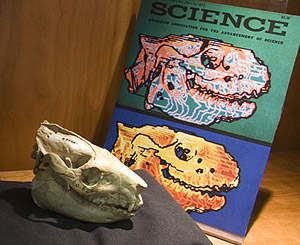
Noninvasive three-dimensional computer imaging of matrix-filled fossil skulls by high-resolution computed tomography

This was the first study to apply newly developed computer imaging techniques originally developed at WUSM for surgical management of craniofacial disorders to the fossil mammal record (Conroy and Vannier 1984). These noninvasive computer imaging techniques generated three-dimensional images of fossil skulls from two-dimensional CT data and allowed paleontologists, for the first time, to electronically “dissect” fossil skulls in different planes by making portions of the skulls, and other obstructing matrix, transparent to reveal previously hidden intracranial morphology. This, and subsequent studies, revolutionized paleontology. These techniques are now used routinely by paleontologists around the world to study the fossil remains of ancient life — from dinosaurs to fossil hominids.
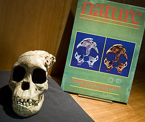
Dental development of the Taung skull from computerized tomography

The discovery in 1924 of the Taung child’s skull from South Africa started an intellectual revolution about human origins that continues unabated to this day (Dart 1925). This skull, dated to about 2.5 million years ago, was the first truly ancient fossil hominid skull ever found on the African continent. Before its discovery, most Victorian-era anthropologists believed that the human lineage first evolved in either Europe or Asia. The Taung discovery changed all that. We now know that fossil hominids existed in Africa nearly 5 million years ago, and that they were confined to that continent until sometime around 1.5 to 2 million years ago when their fossil remains are first found in parts of Europe and Asia. One of the earliest applications of 3D-CT computer imaging to the hominid fossil record was this study of the Taung skull, which showed that its growth and maturational patterns were more apelike than humanlike (Conroy and Vannier 1987).
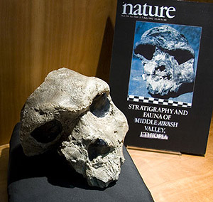
Newly discovered fossil hominid skull from the Afar depression, Ethiopia

The 600,000-year-old cranium from Bodo, Ethiopia, is the oldest and most complete early middle Pleistocene hominid skull from Africa. The cranium was found in 1976 during paleoanthropological explorations in the Afar Desert, Ethiopia, and was described in Nature (Conroy et al. 1978). “Virtual endocast” models created by 3D-CT techniques indicate a brain size of about 1200-1300 cc (Conroy et al. 2000). This discovery proved that by 600,000 years ago, at least one early African hominid species, Homo heidelbergenis, had attained a brain size within the normal range of modern humans.
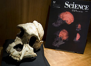
Endocranial capacity in an early hominid cranium from Sterkfontein, South Africa

Two and three-dimensional computer imaging studies showed that this early hominid from South Africa, dated to about 3 million years ago, had a brain size of approximately 515 cc, only about one-third the size of the modern human brain. This was one of the first studies to show that paleoanthropologists could electronically reconstruct missing parts of ancient fossil hominid skulls using 3D-CT techniques and create “virtual endocasts” of such specimens (Conroy et al. 1998).
Bibliography
Conroy, G., Jolly, C., Cramer, D., Kalb, J., 1978. “Newly discovered fossil hominid skull from the Afar depression, Ethiopia.” Nature 275, 67-70.
Conroy, G., Vannier, M., 1987. “Dental development of the Taung skull from computerized tomography.” Nature 329, 625-627.
Conroy, G., Weber, G., Seidler, H., Rechies, W., Nedden, D., Mariam, J., 2000. “Endocranial capacity of the Bodo cranium determined from three-dimensional computed tomography.” Amer. J. Phys. Anthropol. 113, 111-118.
Conroy, G., Weber, G., Seidler, H., Tobias, P., Kane, A., Brunsden, B., 1998. “Endocranial capacity in an early hominid cranium from Sterkfonetin, South Africa.” Science 280, 1730-1731.
Conroy, G., C., Vannier, M.W., 1984. Noninvasive three-dimensional computer imaging of matrix-filled fossil skulls by high-resolution computed tomography. Science 226, 456-458.
Dart, R., 1925. “Australopithecus africanus: the man-ape of South Africa.” Nature 115, 195-199.