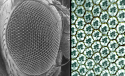
This movie shows a 630 times magnification of the eye of a living fruit fly pupa (the stage just before the fly metamorphoses into an adult). Four cells have been colorized to emphasize their movement as the eye develops. In a competition for a place in the developing eye, the red-colored cell will push between the green-colored cells to contact the underlying primary cell (gray, bean-shaped cell). The movement is controlled by the mutual attraction of proteins Roughest and Hibris on the cells’ surfaces. Both the red and green cells have Roughest protein on their surface. The gray primary cell has Hibris on its surface. After this movie ends, the green cells will have to compete again with other neighbors for a position. In another sector, you can see a blue-colored cell that loses all of its battles and will die. The movie was created by David Larson, a graduate student in the Department of Molecular Biology and Pharmacology.The laws of physics combine with the mutual attraction of two proteins to create the honeycomb pattern of fruit fly eyes, say molecular biologists at Washington University School of Medicine in St. Louis. This same combination of forces forms the delicate filtering structures of the mammalian kidney.
The findings, reported in the June issue of Developmental Cell, provide a new understanding of how individual cells find their niche during organ development. They also mean that the fruit fly eye can now become a fast, inexpensive system for gaining insight into how kidneys develop in mammals and why development sometimes goes awry.
“We’ve challenged scientists who study the development of organs such as eyes and kidneys to think about physics,” says Ross Cagan, Ph.D., associate professor of molecular biology and pharmacology. “In the developing fruit fly eye, we found that cells change shape and move into their proper placement because they want to minimize the free energy of the system.”
Just as molecules of oil floating in water will gather together to exclude water molecules, cells with “sticky” molecules on their surface will gather together in clumps to exclude “non-sticky” cells during organ development. This property of cell adhesion has been previously proposed as a key to moving different cell types into the right positions as developing organs change from an immature, disorganized state to a mature, functional state.
Cagan and his colleague Sujin Bao, Ph.D., research associate in molecular biology and pharmacology, have expanded this principle by showing that cell types possessing two different adhesion molecules, instead of just one, will form a pattern in which one cell type surrounds the other cell type. They found that two proteins, named Roughest and Hibris, play central roles in this process during late stages of development of the fruit fly eye.
“Before the late stages of development, sets of primary cells are surrounded by a disorganized net of support cells,” Cagan says. “But then the cells start producing either Roughest or Hibris on their surfaces, and you see a tight honeycomb pattern of cells take shape.”
Cagan and Bao found that the primary cells in the “holes of the net” express Hibris and the support cells that form the net express Roughest. Roughest and Hibris proteins stick to each other, but they don’t stick to their own kind.
As the proteins appear on the surface of the cells, the laws of physics kick in to move the support cells into positions determined by the energy of attraction. Because Roughest is strongly attracted to Hibris, but not to other Roughest molecules, the support cells are attracted to the surfaces of the primary cells but not to each other. In competition with its neighbors, each Roughest-expressing support cell stretches out as far as it can along a primary cell. Support cells that express less Roughest lose the competition for primary-cell attachment and die off.
At the end of the process, a neat one-cell-thick hexagonal wall of support cells surrounds the primary cells. The repetition of this pattern across the entire fly eye is responsible for the regular honeycomb pattern of the 800 optical units present in the fruit fly compound eye.
“We and others searched for a long time for human equivalents to Roughest and Hibris,” Cagan says. “Surprisingly, they were found in the kidney.”
The equivalent kidney proteins are called Neph1 and Nephrin. They draw together certain kidney-cell junctions in a tight but porous seal that filters urea and other unwanted molecules from the blood vessels within kidney nephrons, structures that filter waste from the blood. Without functioning Neph1 and Nephron, kidneys do not filter properly, leading to neuropathy. Alterations within nephrons also have been linked to hypertension.

The mammalian-kidney versions and the fruit-fly-eye versions of these proteins are fairly specific to their own organs. That is, Neph1 and Nephron are not widely distributed in the mammalian body, and they have no close equivalents in the more primitive kidneys of other kinds of organisms. Roughest and Hibris are found mainly in the late stages of development of the compound eyes of insects related to fruit flies. Interestingly, Neph1 and Nephron are more like Roughest and Hibris than they are like any other protein found in mammals.
“The evolution of these similar proteins in two very distantly related groups of organisms and for these similar purposes suggests that the two systems, the developing kidney and the developing fly eye, used these proteins to solve the same problem—the problem of how to build intricate, fine-structured, tissues from a loose collection of cells,” Cagan says.
“The questions we are asking in the fruit fly—about how cells are sorted—are questions we couldn’t dream of asking by using the mammalian kidney,” Cagan continues. “The fruit fly eye is a much more tractable and faster moving system. When we make discoveries in the fly, such as the roles of Roughest and Hibris, we can then look at the mammalian kidney equivalents, but now in a much more knowledgeable way. Hopefully, we can ‘fast-forward’ research on kidney development.”
Bao S, Cagan R. Preferential adhesion mediated by Hibris and Roughest regulates morphogenesis and patterning in the Drosophila eye. Developmental Cell 2005 June;8:1-11.
Funding from the National Institutes of Health supported this research.
Washington University School of Medicine’s full-time and volunteer faculty physicians also are the medical staff of Barnes-Jewish and St. Louis Children’s hospitals. The School of Medicine is one of the leading medical research, teaching and patient care institutions in the nation, currently ranked third in the nation by U.S. News & World Report. Through its affiliations with Barnes-Jewish and St. Louis Children’s hospitals, the School of Medicine is linked to BJC HealthCare.