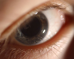
When bright light shines in the eye, the pupil constricts. In dim light, it dilates. Now investigators at Washington University School of Medicine in St. Louis have demonstrated in chickens that a protein called cryptochrome plays a key role in that reflex.
Visual function in vertebrates tends to be regulated by proteins called opsins, and these are the first experiments to suggest that a non-opsin protein plays a dominant role in a visual pathway.
Working with embryonic chicken eyes, Washington University ophthalmology researchers found that cryptochrome allows the pupil to react independently from light-sensitive photoreceptor cells at the back of the eye. The findings are reported in the Oct. 1 issue of the journal Science.

“Light had an effect on the eye without involving the visual pathway,” says principal investigator Russell N. Van Gelder, M.D., Ph.D., an assistant professor of ophthalmology and visual sciences and of molecular biology and pharmacology. “Like a light meter in a camera that measures light levels but doesn’t produce a photo, a nonvisual system in the eye responds to light but doesn’t produce a visual image.”
This new study complements earlier work that had demonstrated the pupils of blind mice still respond to light. Both cryptochrome and another key protein, melanopsin, play important roles in this response. In that nonvisual pathway, melanopsin is thought to play the dominant role in synchronizing the mouse’s internal circadian clock to external light and dark cycles.
In these experiments, however, Van Gelder, and his research team demonstrated that the constriction of the pupil in the chick eye seems to be regulated by cryptochrome rather than melanopsin, Melanopsin is part of the family of proteins, called opsins, that mediate normal visual function. Most opsin proteins are located in the rods and cones of the eye’s retina. But the dissected chick eye had no rods or cones, and its response to light was different than a normal visual response.
In a series of experiments led by first author Daniel C. Tu, a graduate student in Van Gelder’s lab, the researchers looked at the chick eye under various kinds of light and treated it with drugs that disrupt normal, opsin-mediated visual pathways in insects and mammals. None of those experiments disrupted the pupil’s ability to constrict in response to light. But the researchers did get a response when they genetically manipulated the chick eye to reduce production of the cryptochrome protein.
When cryptochrome protein production was blocked by 50 percent, the researchers observed a corresponding 50 percent loss of sensitivity to light. Blocking production of melanopsin had no effect.
“This is still indirect evidence for the involvement of cryptochromes because in the chicken we can’t knock out or overexpress genes like we can in the mouse,” Van Gelder says. “But it does suggest cryptochrome proteins are involved.”
In both mice and chickens, Van Gelder says, it is as if the light meter of the eye is controlling the pupil without vision being involved. In the mouse, the meter is located in the retina and primarily uses melanopsin to do its work with cryptochrome proteins amplifying the signal. In the chick, it is as if the light meter is contained in the pupil itself.
It is not known if the nonvisual pathway seen in the embryonic chick eye is present in mammals. But even if human eyes do not use this particular light-mediated pathway, it still could have broader applications for human health.
“Our hope is to figure out what the building blocks are that make this tissue respond to light in this way and to put them into other cell types,” says Van Gelder. “If we could learn how cryptochrome is making the pupil respond to light, we might be able to make other cells respond to light, even in systems that are not visual. That’s the next phase of our work.”
Tu DC, Batten ML, Palczewski K, Van Gelder RN. Cryptochrome-dependent photoreception in the chick iris. Science, pp. 129-131, Oct. 1, 2004.
This research was funded by grants from the National Eye Institute, Research to Prevent Blindness, the Medical Scientist Training Program of Washington University, the Culpepper Physician Scientist Award of the Rockefeller Brothers Foundation and a grant from the E.K. Bishop Foundation.
The full-time and volunteer faculty of Washington University School of Medicine are the physicians and surgeons of Barnes-Jewish and St. Louis Children’s hospitals. The School of Medicine is one of the leading medical research, teaching and patient care institutions in the nation, currently ranked second in the nation by U.S. News & World Report. Through its affiliations with Barnes-Jewish and St. Louis Children’s hospitals, the School of Medicine is linked to BJC HealthCare.