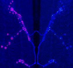
Nerve cells send and receive messages to each other with features akin to “mouths” and “ears:” molecules on the surfaces of cells that send out and receive signals.
Scientists have long suspected that ears deliberately line themselves up opposite mouths on other nerve cells. Now, researchers at Washington University School of Medicine in St. Louis have shown that the theory is valid at the level of the nervous system’s most fundamental unit: individual clusters of mouths and ears.

“We found that receptors don’t develop at random sites — they deliberately develop in areas where they have the best chance of receiving incoming messages,” says Aaron DiAntonio, M.D., Ph.D., assistant professor of molecular biology and pharmacology. “This would be a very useful failsafe mechanism when nerves are making connections with each other during development.”
Although DiAntonio’s results are not directly related to any particular disorder, they add to scientists’ basic understanding of how the nervous system develops, and that body of knowledge may someday help physicians better treat a variety of nervous system disorders.
The findings were published in a recent issue of the journal Current Biology.
Cells communicate by sending chemicals across gaps called synapses. DiAntonio’s group uses a fruit fly model to study neuromuscular junctions, areas where nerve cells enter and exert control over muscle cells.
“All of the known molecules that work in fly neuromuscular junctions are present in the human brain, so we use it as a model to understand how you build this type of synapse,” he explains.
For this study, DiAntonio compared cells from normal fruit flies to those from a mutant strain of fruit fly. The mutants were genetically altered to restrict their ability to make receptors for glutamate, a chemical messenger widely used throughout the nervous system.
His group first showed that nerve cells with fewer glutamate receptors aligned their receptors closely with areas on nearby cells that had the most active sites, features akin to nerve cell mouths that send out the glutamate signal.
In addition to variations in the number of active sites, individual active sites vary in their readiness to pass on a signal. A strong signal is required to get a “sleepy” site to release glutamate, but even very weak signals can cause “alert” sites to release glutamate.

DiAntonio’s team set out to determine whether the mutant cells with fewer receptors tended to align themselves with sleepy active sites or with alert sites. Calcium causes active sites to fire, so the researchers altered levels of calcium surrounding the fruit fly cells. They then stimulated the nerve cells and measured how much of the signal was passed on to receiving cells.
In low calcium conditions, the signal came through just as strong in both mutant and normal cells.
“The alert active sites still fire in low calcium conditions, but the sleepy ones don’t,” DiAntonio explains. “The fact that the mutant cells’ signals came through just as strong in low calcium conditions tells us that their receptors were well-aligned to the alert active sites.”
In high calcium conditions, though, the mutant cells received weaker signals.
“Some of the sleepier active sites start to send signals in those conditions, and the mutant’s reception suffers because it doesn’t have any receptors opposite the sleepier active sites that are now starting to wake up,” DiAntonio explains.
DiAntonio plans to conduct follow-up studies to determine how ears from one nerve cell know to line up near another nerve cell’s mouths.
“Having discovered the phenomenon, we’d like to know what’s making this decision?” he says. “Is it activity in the nerve that triggers receptors to cluster at a particular spot, or are receptors sending feedback to the cell that says, ‘send more receptors this way?'”
Marrus SB, DiAntonio A. Preferential location of glutamate receptors opposite sites of high presynaptic release. Current Biology, June 8, 2004.
Funding from the National Institutes of Health and the W. M. Keck Foundation.
The full-time and volunteer faculty of Washington University School of Medicine are the physicians and surgeons of Barnes-Jewish and St. Louis Children’s hospitals. The School of Medicine is one of the leading medical research, teaching and patient care institutions in the nation, currently ranked second in the nation by U.S. News & World Report. Through its affiliations with Barnes-Jewish and St. Louis Children’s hospitals, the School of Medicine is linked to BJC HealthCare.